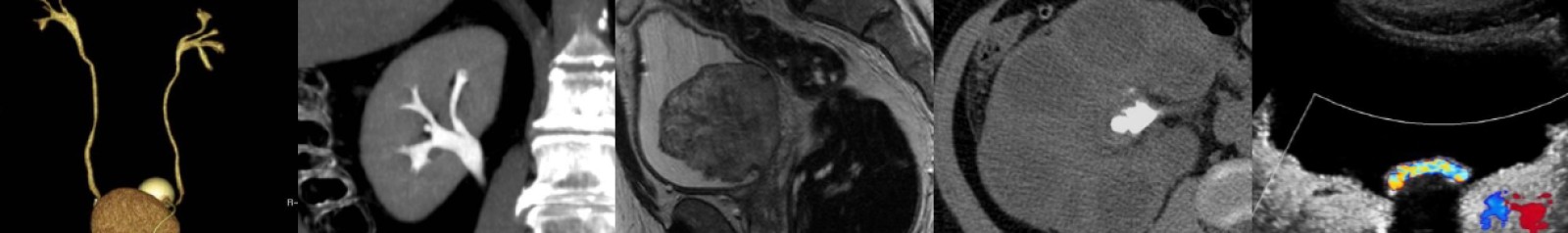
Ultrasound of Endometrioma

In this radiology lecture, we review the ultrasound appearance of endometrioma through three unique cases, including an MRI example.

Key teaching points include:

  • Endometriosis = Ectopic endometrial glands and stroma outside of the uterine cavity. Includes endometriomas, extraovarian implants and adhesions.
  • Endometriomas = Endometriotic cysts within ovary.
  • Endometriosis is seen in about 10% of women of reproductive age.
  • Presentation: Pelvic pain, dysmenorrhea, dyspareunia, infertility.
  • Ultrasound: Diffuse, homogeneous low-level echoes (most specific feature) yielding a ground glass appearance. May have posterior acoustic enhancement.
  • Endometriomas may have peripheral punctate echogenic foci. These foci have no internal vascular flow but can see twinkle artifact.
  • Vascular flow may be present in endometrioma septations.
  • Endometrioma vs. hemorrhagic cyst: Hemorrhagic cysts are acute, usually solitary and unilocular, whereas endometriomas are chronic, sometimes multiple and multilocular.
  • Endometriomas can rarely (1%) undergo malignant transformation into endometrioid carcinoma or clear cell carcinoma.
  • MR is the most specific imaging modality for diagnosis of endometrioma = Specificity 98%.*
  • Homogeneous, T1 “light bulb” bright, T2 dark = “T2 shading.”
  • Surgical treatment: Depends on disease severity from laparoscopic cyst aspiration/cystectomy to hysterectomy/oophorectomy.
  • Medical management may be attempted: Oral contraceptives, GnRH agonists

*Reference: Togashi K, Nishimura K, Kimura I, et al. Endometrial cysts: Diagnosis with MR imaging. Radiology. 1991;180:73-78.

To learn more about the Samsung RS85 Prestige ultrasound system, please visit: https://www.bostonimaging.com/rs85-prestige-ultrasound-system-4

Click the YouTube Community tab or follow on social media for bonus teaching material posted throughout the week!

Instagram: https://www.instagram.com/radiologistHQ/
Facebook: https://www.facebook.com/radiologistHeadQuarters/
Twitter: https://twitter.com/radiologistHQ


Ultrasound of Testicular Torsion

In this radiology lecture, we review the ultrasound appearance of testicular torsion through three unique cases.

Key teaching points include:

  • Torsion occurs when spermatic cord twists and cuts off blood supply to the testis.
  • Bell-clapper deformity most common etiology: Abnormally high attachment of tunica vaginalis allowing spermatic cord rotation and testicular torsion (intravaginal).
  • Torsion has a bimodal distribution: First year of life (extravaginal), adolescents/young adults (intravaginal).
  • “Whirlpool” sign: Eddy swirl of coiled spermatic cord superior to testis, highly specific but less commonly seen than redundant spermatic cord.
  • Redundant spermatic cord AKA boggy pseudomass, torsion knot, epididymal-cord complex and should be avascular or only minimally vascular (unlike paratesticular neoplasm or acute epididymitis).
  • Testicles normally lie vertically, but horizontal or oblique (diagonal) lie suspicious for torsion.
  • Testicular enlargement, reactive hydrocele and scrotal skin thickening are secondary findings of torsion.
  • Marked testicular heterogeneity = Late torsion and nonviability/necrosis, more likely after 24 hours of symptoms.
  • Treatment: Detorsion and orchiopexy if salvageable, orchiectomy if not.

Reference: Bandarkar AN, Blask AR. Testicular torsion with preserved flow: Key sonographic features and value-added approach to diagnosis. Pediatric Radiology (2018) 48:735–744.

To learn more about the Samsung RS85 Prestige ultrasound system, please visit: https://www.bostonimaging.com/rs85-prestige-ultrasound-system-4

Click the YouTube Community tab or follow on social media for bonus teaching material posted throughout the week!

Instagram: https://www.instagram.com/radiologistHQ/
Facebook: https://www.facebook.com/radiologistHeadQuarters/
Twitter: https://twitter.com/radiologistHQ

Ultrasound & CT of Renal Oncocytoma

In this radiology lecture, the ultrasound and CT appearance of renal oncocytoma is revealed.

Key teaching points include:

  • Oncocytomas are benign, solid tumors.
  • 13% patients have multiple oncocytomas, and 1/3rd have concurrent renal cell carcinoma.
  • Central stellate nonenhancing scar only seen in 1/3rd of cases, and more commonly in larger tumors.
  • Spoke-wheel angiographic pattern may be present, best visualized on ultrasound using microvascular flow, AKA superb microvascular imaging.
  • Often not possible to differentiate from renal cell carcinoma (RCC) with imaging.
  • Both oncocytoma and RCC can have central scar and/or spoke-wheel angiographic pattern.
  • Kim et al.* found segmental enhancement inversion (corticomedullary phase/early excretory phase) characteristic for oncocytoma, but subsequent studies have shown inconsistent results.
  • When evaluating a renal mass, if only have postcontrast and 15-minute delayed phase images, mass deenhancement of 15 HU or more suggests a solid mass, whereas no change is more consistent with a hyperattenuating cyst.**

*Kim JI, Cho JY, Moon KC, et al. Segmental enhancement inversion at biphasic multidetector CT: Characteristic finding of small renal oncocytoma. Radiology 2009;252(2):441–448.

**Macari M, Bosniak MA. Delayed CT to evaluate renal masses incidentally discovered at contrast-enhanced CT: Demonstration of vascularity with deenhancement. Radiology 1999;213:674-680.

To learn more about the Samsung RS85 Prestige ultrasound system, please visit: https://www.bostonimaging.com/rs85-prestige-ultrasound-system-4

Click the YouTube Community tab or follow on social media for bonus teaching material posted throughout the week!

Instagram: https://www.instagram.com/radiologistHQ/
Facebook: https://www.facebook.com/radiologistHeadQuarters/
Twitter: https://twitter.com/radiologistHQ

Case of the Week: Ovarian Mucinous Cystadenocarcinoma (Ultrasound & MRI)

In this radiology lecture, we reveal the imaging appearance of mucinous cystadenocarcinoma of the ovary and explain differentiating features from serous cystadenocarcinoma.

Key points include:

  • A rare type of malignant ovarian epithelial tumor.
  • Often large at presentation, may be enormous.
  • Almost always multilocular.
  • Mucinous, proteinaceous and hemorrhagic material within loculi.
  • US: Scattered low-level echoes.
  • MRI: “Stained glass” appearance = Variable T1/T2 signal. Thick mucin = T1/T2 hyperintense.
  • Irregular, thick septations and solid components with internal vascularity and enhancement allow differentiation from mucinous cystadenoma.

Click the YouTube Community tab or follow on social media for bonus teaching material posted throughout the week!

Instagram: https://www.instagram.com/radiologistHQ/
Facebook: https://www.facebook.com/radiologistHeadQuarters/
Twitter: https://twitter.com/radiologistHQ

Ultrasound of Complete Molar Pregnancy

In this radiology lecture, the ultrasound appearance of complete molar pregnancy is revealed.

Key points include:

  • AKA hydatiform mole = Most common form of gestational trophoblastic disease.
  • Gestational trophoblastic neoplasia (GTN) less common = Invasive mole and choriocarcinoma.
  • Approximately 1/1,000 pregnancies is a molar pregnancy.
  • Most common in females under age 20 and over age 35.
  • Two types of molar pregnancy: Complete (most common) and partial.
  • Complete: Diploid (paternal DNA only), no fetus, more likely to be complicated by GTN.
  • Partial: Triploid (maternal and paternal DNA), abnormal fetus or fetal parts, harder to diagnose.
  • Complete hydatiform mole presentation: Vaginal bleeding, enlarged uterus inconsistent with dates, hyperemesis. Markedly elevated β-hCG level (variable for partial molar pregnancies).
  • Large theca lutein cysts due to ovarian stimulation from elevated β-hCG, but uncommon.
  • US: Heterogeneous, echogenic mass (“snowstorm” appearance), small anechoic cystic spaces (“cluster of grapes”) = hydropic chorionic villi.
  • Treatment: Dilation & curettage. β-hCG levels monitored until no longer detectable to confirm no residual disease.

To learn more about the Samsung RS85 Prestige ultrasound system, please visit:   https://www.bostonimaging.com/rs85-prestige-ultrasound-system-4

Click the YouTube Community tab or follow on social media for bonus teaching material posted throughout the week!

Instagram: https://www.instagram.com/radiologistHQ/
Facebook: https://www.facebook.com/radiologistHeadQuarters/
Twitter: https://twitter.com/radiologistHQ

Case of the Week: Wandering Spleen (CT)

In this radiology lecture, we discuss the CT appearance of wandering spleen!

Key points include:

  • Extremely rare, usually between 20-40 years of age, more common in females.
  • Splenic mobility due to congenital or acquired abnormality of the normal peritoneal attachments/suspensory ligaments.
  • Splenic migration to lower abdomen/pelvis, may develop long vascular pedicle.
  • Twisting of pedicle can lead to splenic ischemia and infarction if not promptly treated.
  • Variable clinical presentation, patients often become symptomatic if torsion of pedicle occurs: Intermittent colicky pain, vague abdominal discomfort, abdominal mass, acute abdomen.
  • Treatment: Surgical detorsion and fixation of spleen (splenopexy), splenectomy may be required in setting of infarction.

Click the YouTube Community tab or follow on social media for bonus teaching material posted throughout the week!

Instagram: https://www.instagram.com/radiologistHQ/
Facebook: https://www.facebook.com/radiologistHeadQuarters/
Twitter: https://twitter.com/radiologistHQ

5 Cases in 5 Minutes: Musculoskeletal #4

Quiz yourself with this week’s interactive video lecture as we present a total of 5 interesting musculoskeletal radiology cases followed by a diagnosis reveal and key teaching points after each case, all in just a few minutes!

Click the YouTube Community tab or follow on social media for bonus teaching material posted throughout the week!

Instagram: https://www.instagram.com/radiologistHQ/
Facebook: https://www.facebook.com/radiologistHeadQuarters/
Twitter: https://twitter.com/radiologistHQ

Case of the Week: Septate Uterus (MRI)

In this radiology lecture, we discuss the MRI appearance of septate uterus, and explain how to differentiate from other uterine anomalies.

Key points include:

  • Most common müllerian duct anomaly (55%): Septal reabsorption abnormality.
  • Ultrasound and MRI provide assessment of external uterine contour and presence of renal anomalies.
  • Hysterosalpingogram of limited value, cannot reliably differentiate between subtypes.
  • On MRI, uterine fundus is typically convex or minimally indented: Fundal cleft less than 1 cm.
  • Midline septum of variable length, may be muscular or fibrous.
  • Important to differentiate type of septum as may alter surgical approach.
  • Compared to bicornuate uterus, higher incidence of reproductive complications (miscarriage).
  • Treatment: Resection of septum if recurrent fetal loss.

Click the YouTube Community tab or follow on social media for bonus teaching material posted throughout the week!

Instagram: https://www.instagram.com/radiologistHQ/
Facebook: https://www.facebook.com/radiologistHeadQuarters/
Twitter: https://twitter.com/radiologistHQ

Ultrasound of Ovarian Dermoid Cyst

In this radiology lecture, we discuss the ultrasound appearance of ovarian dermoid cyst, including the rarely seen but highly specific “floating sphere” sign!

Key points include

  • AKA mature cystic teratoma.
  • Most common ovarian neoplasm.
  • Benign, mean age 30.
  • 10% bilateral.
  • Mature tissue from ≥2 embryonic germ cell layers: Sebaceous material, hair follicles, skin derivatives, fat, muscle, bone, and other tissues lined by squamous epithelium.
  • Specificity of US diagnosis 94-100%.
  • MRI for changing morphology on f/u and for postmenopausal patients.
  • Ultrasound findings: Floating echogenic spherical structures = “Floating sphere” sign (uncommon but pathognomonic), hyperechoic component with acoustic shadowing (Rokitanksy nodule), hyperechoic lines and dots, fat-fluid levels, diffuse or regional bright echoes.
  • Most common complication: Ovarian torsion.
  • Rare complications: Rupture, infection, malignant transformation, hormone secretion, anti-NMDA receptor encephalitis.

To learn more about the Samsung RS85 Prestige ultrasound system, please visit: https://www.bostonimaging.com/rs85-prestige-ultrasound-system-4

Click the YouTube Community tab or follow on social media for bonus teaching material posted throughout the week!

Instagram: https://www.instagram.com/radiologistHQ/
Facebook: https://www.facebook.com/radiologistHeadQuarters/
Twitter: https://twitter.com/radiologistHQ

Case of the Week: Retroperitoneal Fibrosis (Ultrasound & CT)

In this radiology lecture, we discuss the ultrasound and CT appearance of retroperitoneal fibrosis.

Key points include:

  • Most cases (70%) are idiopathic = Ormond disease.
  • Nonspecific symptoms depending on involved structures: Malaise, weight loss, low-grade fever.
  • Ureteral entrapment: Obstructive uropathy or renal failure, may see medial deviation of middle third of ureters with hydronephrosis.
  • Venous entrapment: Lower extremity edema, deep venous thrombosis.
  • CT: Soft tissue mass anterolateral to aorta with posterior sparing.
  • DDx: Retroperitoneal Lymphoma will Lift the aorta.
  • MRI: Low T1/T2 signal when inactive, T2 bright with early enhancement when active inflammation.
  • PET/CT: Avid when metabolically active, may aid in identifying appropriate biopsy sites.

Click the YouTube Community tab or follow on social media for bonus teaching material posted throughout the week!

Instagram: https://www.instagram.com/radiologistHQ/
Facebook: https://www.facebook.com/radiologistHeadQuarters/
Twitter: https://twitter.com/radiologistHQ

1 2 3 4 5 6 8