Case of the Week: Ovarian Mucinous Cystadenocarcinoma (Ultrasound & MRI)
In this radiology lecture, we reveal the imaging appearance of mucinous cystadenocarcinoma of the ovary and explain differentiating features from serous cystadenocarcinoma.
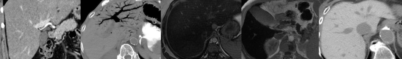
Key points include:
- A rare type of malignant ovarian epithelial tumor.
- Often large at presentation, may be enormous.
- Almost always multilocular.
- Mucinous, proteinaceous and hemorrhagic material within loculi.
- US: Scattered low-level echoes.
- MRI: “Stained glass” appearance = Variable T1/T2 signal. Thick mucin = T1/T2 hyperintense.
- Irregular, thick septations and solid components with internal vascularity and enhancement allow differentiation from mucinous cystadenoma.
Click the YouTube Community tab or follow on social media for bonus teaching material posted throughout the week!
Instagram: https://www.instagram.com/radiologistHQ/
Facebook: https://www.facebook.com/radiologistHeadQuarters/
Twitter: https://twitter.com/radiologistHQ
Podcast: Play in new window | Download
Subscribe: Email