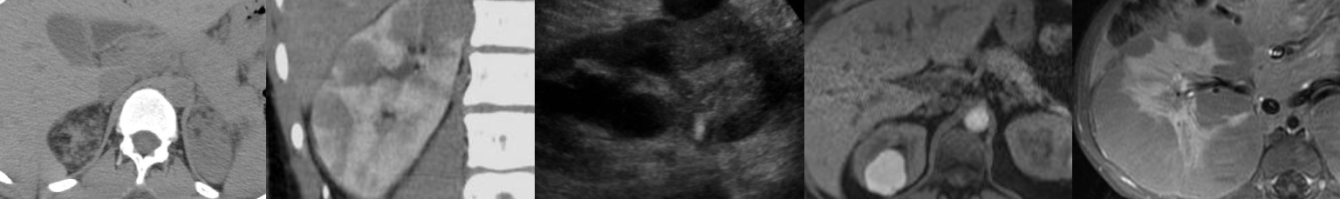
Introduction to Multiphase CT & MRI of the Liver

In this video lecture, we review the appearance of the liver on multiphase CT & MRI. A basic approach to image interpretation is presented with pitfalls to avoid.

Key points include:

  • The three major liver postcontrast phases include the late hepatic arterial phase, portal venous phase, and delayed/equilibrium phases.
  • The hepatic artery enhances first, followed by the portal veins, then the hepatic veins along with the hepatic parenchyma.
  • An ideal late hepatic arterial phase sequence will have both hepatic artery and portal vein enhancement with no hepatic vein enhancement.
  • The late hepatic arterial phase occurs at about the same time as the corticomedullary phase, enteric phase, pancreatic phase, and splenic arciform enhancement phase.
  • The early arterial phase of an angiographic CT is NOT the same as the late hepatic arterial phase of a liver protocol study and may be too early to adequately assess hypervascular liver lesions.
  • “MRI CT” is a handy mnemonic for hypervascular liver metastases, lesions that will be best detected on a hepatic arterial phase series.
  • Portal venous phase images will have portal vein and hepatic vein enhancement, as well as liver parenchymal enhancement.
  • Hypovascular hepatic metastases (GI tract, pancreas) are usually best detected on the portal venous phase.
  • Delayed/equilibrium phase images allow detection of intralesional contrast washout and delayed capsular enhancement typical of hepatocellular carcinoma, as well as evaluation of delayed enhancement as seen with hemangiomas and intrahepatic cholangiocarcinoma.
  • CT has better spatial resolution, but MRI has better contrast resolution and is therefore superior to CT in the characterization of liver masses.
  • Pre- and postcontrast MRI sequences are typically obtained as a special T1 sequence known as a spoiled 3D gradient echo variant with fat saturation.

Hepatic Hemangioma: Pitfalls & Mimics, Part I

In this video lecture, we discuss tips and tricks to diagnose everybody’s favorite hepatic tumor on CT, MRI and ultrasound.

Key points include:

  • Hemangioma is the most common benign hepatic tumor, and it is more common in females.
  • These tumors are usually asymptomatic and typically require no treatment, but can rarely cause pain, rupture if large, or cause Kasabach-Merritt syndrome.
  • On nonenhanced CT, hemangiomas will be hypodense to liver parenchyma and homogeneously isodense to the blood pool.
  • There are three major enhancement patterns for typical hemangiomas, and all patterns will show persistent delayed enhancement without contrast washout.
  • Peripheral, nodular, interrupted enhancement with gradual centripetal progression to uniform enhancement is the most common pattern.
  • Smaller lesions (less than 1-2 cm) can have immediate uniform enhancement and appear flash-filling.
  • Larger hemangiomas may have a central scar that does not enhance.
  • MRI is highly specific in the diagnosis of hemangioma.
  • On MRI, hemangiomas will appear T1 hypointense and T2 hyperintense to liver parenchyma, or (perhaps more importantly) T1 isointense to the blood pool and T2 hyperintense to the spleen.
  • Hemangiomas usually do not show restricted diffusion.
  • If present, the central scar of hemangioma will appear T1 hypointense and T2 hyperintense on MRI.
  • Additional liver masses that may have a central scar include focal nodular hyperplasia, fibrolamellar hepatocellular carcinoma, cholangiocarcinoma, and hepatocellular carcinoma.
  • On ultrasound, hemangiomas are usually uniformly echogenic.
  • 40% of hemangiomas can have a “reverse target” appearance with an echogenic periphery and hypoechoic center.
  • Hemangiomas usually have no color Doppler flow on ultrasound, but they may occasionally exhibit mild flow.

Hepatic Hemangioma: Pitfalls & Mimics, Part II

In this video lecture, we focus on variants and malignant mimics of hemangioma and discuss how to characterize these masses on ultrasound, CT and MRI.

Key points include:

  • The ultrasound “target” sign is typical for hepatic metastases and appears as a lesion with a hypoechoic periphery and echogenic center.
  • Hemangiomas in a fatty liver may appear hypoechoic and mimic a more serious tumor but can be definitively characterized with MRI.
  • Sclerosed or hyalinized hemangiomas contain fibrous tissue and therefore have variable enhancement and diminished T2 hyperintensity.
  • Hemangiomas can be associated with arterioportal shunts and may be surrounded by areas of transient hepatic enhancement difference (THED) and peritumoral sparing of fatty infiltration.
  • Differential diagnostic considerations for hepatic hemangioma include hypervascular liver metastases (including melanoma and pancreatic neuroendocrine tumor), intrahepatic mass-forming cholangiocarcinoma, and hepatic angiosarcoma.
  • Irregular rim enhancement is not the same as interrupted peripheral nodular enhancement and should raise suspicion for malignancy in a solid mass.
  • Metastatic tumors may progressively fill in and mimic hemangioma, but often show less pronounced “evil grey” T2 hyperintensity isointense to spleen, restrict diffusion, and have atypical enhancement.
  • Mass-forming, peripheral intrahepatic cholangiocarcinoma can mimic hemangioma as both tumors demonstrate gradual progressive enhancement, but cholangiocarcinoma enhancement will be heterogeneous and not usually isointense to blood pool.
  • Intrahepatic cholangiocarcinoma is heterogeneously T2 hyperintense often with central hypointensity and overlying capsular retraction but may not have associated biliary ductal dilatation.