In the conclusion of this introductory series, we discuss the basics of computed tomography (CT) urography, bladder and urethral diverticula, and scrotal pathology including varicocele, epididymo-orchitis, and testicular neoplasm.
Topics include:
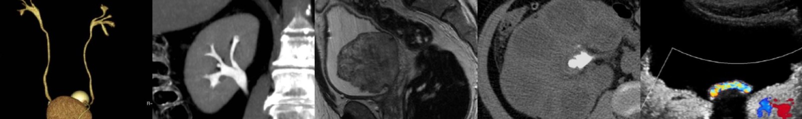
- CT urography post-processing techniques: maximum intensity projection (MIP) and 3D volume-rendered (VR) imaging.
- Appearance of transitional cell carcinoma in the ureters and bladder on excretory phase CT mages.
- Papillary necrosis.
- Weigert-Meyer rule for duplicated collecting systems and tips to remember it.
- Urethral diverticula appearance on magnetic resonance imaging (MRI) and voiding cystourography (VCUG).
- Scrotal varicocele and the importance of recognizing the implications of right-sided versus left-sided.
- Acute scrotal pathology: testicular torsion and epididymo-orchitis.
- Differentiating seminomatous and non-seminomatous testicular neoplasms via ultrasound.
Podcast: Play in new window | Download
Subscribe: Email