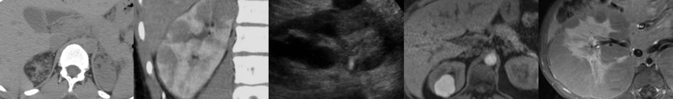
Non-masslike Adrenal Abnormalities

In this video lecture, we discuss the normal imaging appearance of the adrenal glands, as well as multiple common and rare non-masslike adrenal abnormalities on CT and MRI.

Key points include:

  • The adrenal glands normally have an inverted “V” or “Y” configuration.
  • A linear, disk-like adrenal gland or “pancake” adrenal gland indicates congenital malposition or absence of the ipsilateral kidney.
  • Adrenal hyperenhancement raises suspicion for developing shock and may signal a therapeutic window to act upon.
  • Adrenal calcifications can be caused by prior hemorrhage, infection, tumors and metabolic syndromes.
  • On MRI, T1-weighted in-phase GRE images can be helpful in identifying calcification due to dark blooming caused by susceptibility artifact.
  • Adrenal atrophy may indicate adrenal insufficiency.
  • Adrenal hyperplasia is identified when adrenal limbs measure greater than 10 mm in thickness and may or may not be hormonally functioning.
  • Nonhemorrhagic adrenal infarction appears as a nonenhancing, edematous adrenal gland with surrounding stranding and restricted diffusion.
  • Adrenal infarction can be seen in hypercoagulable states, antiphospholipid-antibody syndrome and pregnancy.

Imaging of Adrenal Adenomas & Incidentalomas

In this video lecture, we discuss the imaging appearance of lipid-rich and lipid-poor adrenal adenomas, explain the CT washout calculation, and review the choice of CT vs. MRI for the evaluation of adrenal nodules. Also, we compare the 2017 ACR (American College of Radiologists) Incidental Findings Committee recommendations to the AACE/AAES (American Association of Clinical Endocrinologists and American Association of Endocrine Surgeons) medical guidelines in the evaluation of adrenal incidentalomas.

Key points include:

  • 70% of adrenal adenomas will be lipid-rich with a low-density of 10 or less Hounsfield units (HU) on CT.
  • Adrenal nodules with greater than 60% enhancement washout and greater than 40% relative washout are consistent with adenomas.
  • Using chemical shift, MRI may be helpful in characterizing a mass which is indeterminate on non-contrast CT, particularly if the non-contrast density is less than 20-30 HU.
  • If the non-contrast density of an adrenal nodule is greater than 20-30 HU, MRI should be avoided, as washout CT is typically the test of choice.
  • While the AACE/AAES and ACR follow-up algorithms for adrenal nodules differ in many respects, they agree that surgical resection should be considered for adrenal masses measuring 4 cm or more in size, and that biochemical evaluation should at least be considered for most adrenal masses.

Adrenal Hemorrhage

In this video lecture, we discuss the imaging appearance of adrenal hemorrhage on CT, MRI and ultrasound. Causes of adrenal hemorrhage will also be reviewed. 

Key points include:

  • On CT scan, adrenal hemorrhage typically appears as a nonenhancing round or oval hyperdense mass with density of 50-90 HU. 
  • MRI is the most sensitive and specific modality for diagnosing adrenal hemorrhage. 
  • The “high signal intensity rim” rim sign seen on T1-weighted images is characteristic of subacute adrenal hemorrhage. 
  • Adrenal hemorrhage may appear solid or cystic on ultrasound depending on the age of hemorrhage.
  • Adrenal hemorrhage is more common in neonates than in children and adults and is the most common adrenal mass in infancy.
  • Trauma is the most common cause and is usually unilateral and right-sided. 
  • Atraumatic adrenal hemorrhage is usually unilateral.

Adrenal Cysts and Myelolipomas

In this video lecture, we discuss the imaging appearance of adrenal cysts and myelolipomas, as well as differential diagnostic considerations.

Key points include:

  • Adrenal cysts typically have density of simple fluid ranging from 20 to -10 Hounsfield units (HU).
  • Adrenal pseudocysts may be symptomatic, may have a complicated appearance and may have peripheral calcification.
  • The “claw” sign can be helpful in differentiating adrenal from renal masses.
  • Gastric diverticula can mimic adrenal cysts but can be differentiated by identifying communication of gastric diverticulum with adjacent stomach, as well as the presence of oral contrast and/or gas with the diverticulum.
  • Adrenal myelolipomas are benign neoplasms that have varying degrees of macroscopic fat and hematopoietic soft tissue elements.
  • Fat density within myelolipomas on CT ranges from -30 to -90 HU.
  • Up to 30% of myelolipomas contain small calcifications.
  • Unlike myelolipomas, malignant retroperitoneal sarcomas are usually ill-defined and displace or invade adjacent structures.
  • Because myelolipomas contain macroscopic fat, they show greater signal loss (darkening) on fat-saturated images compared to T1 opposed-phase chemical shift images.
  • Conversely, lipid-rich adrenal adenomas contain microscopic fat (also known as intracellular, intracytoplasmic, or intravoxel fat) and therefore show greater signal loss on T1 opposed-phase images than on fat-saturated images.

Imaging of Pheochromocytoma

In this video lecture, we discuss the diagnosis and imaging appearance of pheochromocytoma on ultrasound, CT, MRI, and nuclear medicine studies.

Key points include:

  • Pheochromocytoma is a biochemical diagnosis.
  • Symptoms are variable, and patients may present with headache, palpitations, sweating, hypertension or be asymptomatic.
  • Most cases are sporadic with mean age of presentation in the 40s.
  • Hereditary pheochromocytoma can be seen in the setting of Von Hippel-Lindau disease, multiple endocrine neoplasia (MEN) type II, neurofibromatosis type I, and pheochromocytoma-paraganglioma syndromes associated with succinate dehydrogenase gene mutations.
  • Pheochromocytoma is the “10%” tumor, as approximately 10% are bilateral, extra-adrenal, contain calcification, occur in children, or are malignant.
  • Most pheochromocytomas are solid, but they can also appear as mixed cystic and solid with varying degrees of complexity.
  • The ultrasound appearance is nonspecific, and tumors may contain echogenic hemorrhage or be hypervascular on Doppler imaging.
  • Any mass that enhances to a density greater than 110 HU on an arterial phase CT scan should raise suspicion for pheochromocytoma.
  • On CT, most pheochromocytomas will have noncontrast density greater than 10 HU and absolute washout less than 60%, findings similar to adrenal metastases and adrenocortical carcinoma.
  • However, pheochromocytoma can rarely have noncontrast density less than 10 HU and absolute washout greater than 60%, thus mimicking adrenal adenoma.
  • Studies have shown that pheochromocytoma is not a contraindication to modern nonionic IV contrast media.
  • On MRI, the “light bulb” T2 bright appearance classically associated with pheochromocytoma may or may not be present.
  • If intratumoral hemorrhage is present, pheochromocytomas may show T1 hyperintensity or susceptibility artifact on T1-weighted in-phase images depending on the age of blood.
  • Pheochromocytomas usually do not show signal dropout on T1-weighted opposed-phase images but rarely do and can mimic adrenal adenomas.
  • MIBG scans have nearly 100% specificity for pheochromocytoma and are useful to confirm clinically suspected pheochromocytoma and for detection of metastatic disease.
  • Extra-adrenal pheochromocytomas (paragangliomas) usually occur in abdomen, typically at the organ of Zuckerkandl or in the retroperitoneum.

Malignant Adrenal Masses

In this video lecture, we discuss the diagnosis and imaging appearance of malignant adrenal masses: adrenal metastases including collision tumor, adrenocortical carcinoma and adrenal lymphoma.

Key points include:

  • Adrenal metastases are the most common malignant lesion involving the adrenal gland.
  • Lung carcinoma is the most common primary malignancy to metastasize to the adrenal glands.
  • Adrenal metastases are often bilateral and greater than 3 cm in size.
  • When malignant adrenal lesions are compared to adenomas, SUV cutoff of 3.1 has a 99% negative predictive value.
  • Adrenal-to-liver SUV ratio cutoff value of 1.4 has a specificity of 100% in differentiating adrenal adenomas and metastases.
  • Collision tumors are two histologically distinct tumors that abut or are near each other in the adrenal gland, and PET/CT is the best way to characterize these lesions without biopsy.
  • An enlarging defect within adrenal signal dropout on T1-weighted opposed-phase GRE images is suspicious for a metastatic collision tumor abutting a lipid-rich adrenal adenoma.
  • Renal cell carcinoma metastases can be slow growing and occur many years after the initial tumor presentation.
  • Adrenocortical carcinoma has a bimodal age distribution, may be hormonally functioning and has a poor prognosis.
  • Adrenocortical carcinoma usually presents as a large (greater than 6 cm) mass with internal hemorrhage, necrosis and sometimes calcification.
  • Venous invasion is common with adrenocortical carcinoma.
  • Adrenal lymphoma will be round or adreniform in shape and frequently shows restricted diffusion, a feature that can be helpful in differentiating from adrenal hyperplasia.
  • Diffuse large B-cell lymphoma is the most common type of adrenal lymphoma, and patients usually present with B-cell symptoms and/or adrenal insufficiency.
  • Adrenal lymphoma is usually bilateral and may invade the adjacent kidney(s).