Adrenal Hemorrhage
In this video lecture, we discuss the imaging appearance of adrenal hemorrhage on CT, MRI and ultrasound. Causes of adrenal hemorrhage will also be reviewed.
Key points include:
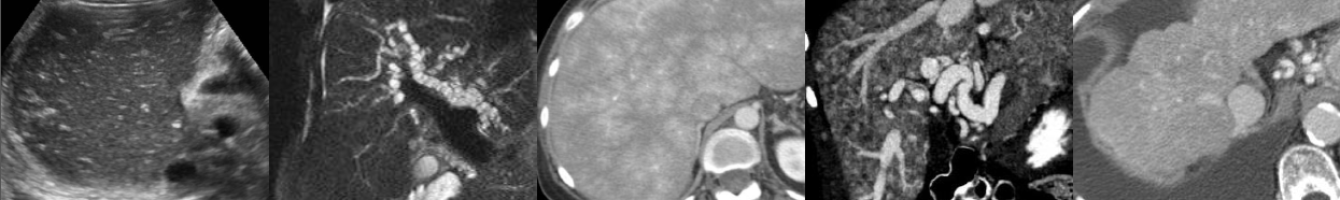
- On CT scan, adrenal hemorrhage typically appears as a nonenhancing round or oval hyperdense mass with density of 50-90 HU.
- MRI is the most sensitive and specific modality for diagnosing adrenal hemorrhage.
- The “high signal intensity rim” rim sign seen on T1-weighted images is characteristic of subacute adrenal hemorrhage.
- Adrenal hemorrhage may appear solid or cystic on ultrasound depending on the age of hemorrhage.
- Adrenal hemorrhage is more common in neonates than in children and adults and is the most common adrenal mass in infancy.
- Trauma is the most common cause and is usually unilateral and right-sided.
- Atraumatic adrenal hemorrhage is usually unilateral.
Podcast: Play in new window | Download
Subscribe: Email