Malignant Adrenal Masses
In this video lecture, we discuss the diagnosis and imaging appearance of malignant adrenal masses: adrenal metastases including collision tumor, adrenocortical carcinoma and adrenal lymphoma.
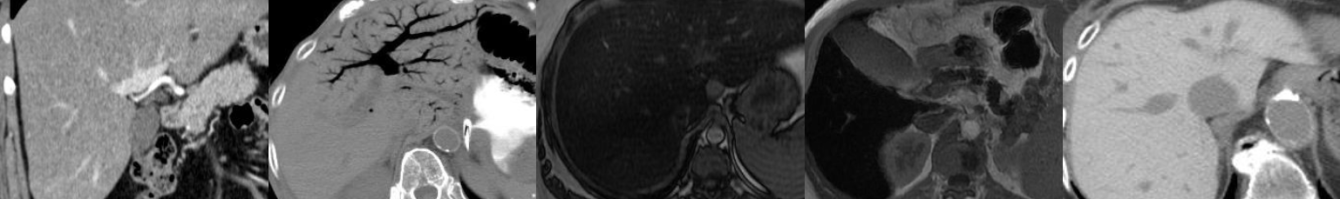
Key points include:
- Adrenal metastases are the most common malignant lesion involving the adrenal gland.
- Lung carcinoma is the most common primary malignancy to metastasize to the adrenal glands.
- Adrenal metastases are often bilateral and greater than 3 cm in size.
- When malignant adrenal lesions are compared to adenomas, SUV cutoff of 3.1 has a 99% negative predictive value.
- Adrenal-to-liver SUV ratio cutoff value of 1.4 has a specificity of 100% in differentiating adrenal adenomas and metastases.
- Collision tumors are two histologically distinct tumors that abut or are near each other in the adrenal gland, and PET/CT is the best way to characterize these lesions without biopsy.
- An enlarging defect within adrenal signal dropout on T1-weighted opposed-phase GRE images is suspicious for a metastatic collision tumor abutting a lipid-rich adrenal adenoma.
- Renal cell carcinoma metastases can be slow growing and occur many years after the initial tumor presentation.
- Adrenocortical carcinoma has a bimodal age distribution, may be hormonally functioning and has a poor prognosis.
- Adrenocortical carcinoma usually presents as a large (greater than 6 cm) mass with internal hemorrhage, necrosis and sometimes calcification.
- Venous invasion is common with adrenocortical carcinoma.
- Adrenal lymphoma will be round or adreniform in shape and frequently shows restricted diffusion, a feature that can be helpful in differentiating from adrenal hyperplasia.
- Diffuse large B-cell lymphoma is the most common type of adrenal lymphoma, and patients usually present with B-cell symptoms and/or adrenal insufficiency.
- Adrenal lymphoma is usually bilateral and may invade the adjacent kidney(s).
Podcast: Play in new window | Download
Subscribe: Email