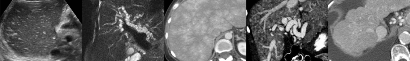
Ultrasound of Complete Molar Pregnancy
In this radiology lecture, the ultrasound appearance of complete molar pregnancy is revealed.
Key points include:
- AKA hydatiform mole = Most common form of gestational trophoblastic disease.
- Gestational trophoblastic neoplasia (GTN) less common = Invasive mole and choriocarcinoma.
- Approximately 1/1,000 pregnancies is a molar pregnancy.
- Most common in females under age 20 and over age 35.
- Two types of molar pregnancy: Complete (most common) and partial.
- Complete: Diploid (paternal DNA only), no fetus, more likely to be complicated by GTN.
- Partial: Triploid (maternal and paternal DNA), abnormal fetus or fetal parts, harder to diagnose.
- Complete hydatiform mole presentation: Vaginal bleeding, enlarged uterus inconsistent with dates, hyperemesis. Markedly elevated β-hCG level (variable for partial molar pregnancies).
- Large theca lutein cysts due to ovarian stimulation from elevated β-hCG, but uncommon.
- US: Heterogeneous, echogenic mass (“snowstorm” appearance), small anechoic cystic spaces (“cluster of grapes”) = hydropic chorionic villi.
- Treatment: Dilation & curettage. β-hCG levels monitored until no longer detectable to confirm no residual disease.
To learn more about the Samsung RS85 Prestige ultrasound system, please visit: https://www.bostonimaging.com/rs85-prestige-ultrasound-system-4
Click the YouTube Community tab or follow on social media for bonus teaching material posted throughout the week!
Instagram: https://www.instagram.com/radiologistHQ/
Facebook: https://www.facebook.com/radiologistHeadQuarters/
Twitter: https://twitter.com/radiologistHQ
Podcast: Play in new window | Download
Subscribe: Email