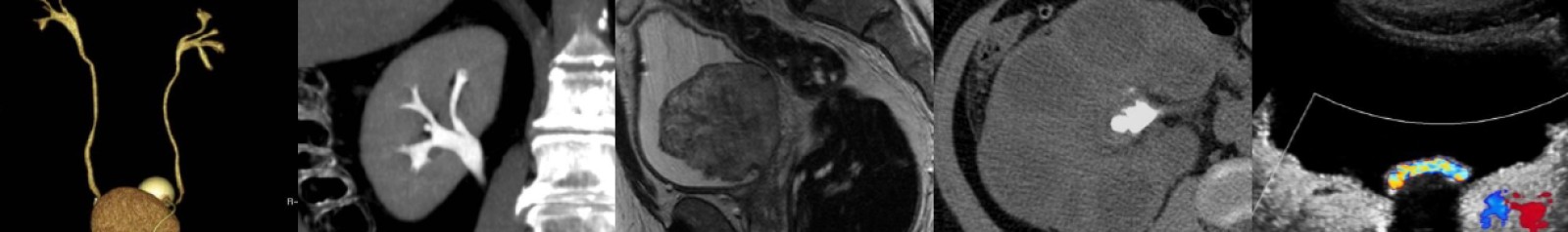
Case of the Week: Pulmonary Infarction (X-ray & CT)
In this radiology lecture, we discuss the chest x-ray and CT appearance of pulmonary infarction in the setting of acute pulmonary embolism.
Key points include:
- Uncommon complication of pulmonary embolism.
- Most common in right lung.
- Risk of infarction increases with large clot burden.
- Typically wedge-shaped, peripheral consolidation with no air bronchograms (Hampton hump).
- However, may not be wedge-shaped, and not all wedge-shaped opacities will be infarcts in the setting of pulmonary embolism.
- “Bubbly” consolidation containing rounded, central lucencies: Most specific finding of infarct* and represents a combination of infarcted, necrotic lung and adjacent viable, aerated lung.
- “Vessel” sign: Enlarged vessel leading to apex of a wedge-shaped opacity. Vessel is dilated due to the presence of intraluminal thrombus or distal obstruction.
*Revel MP, Triki R, Chatellier G, et al. Is it possible to recognize pulmonary infarction on multisection CT images? Radiology. 2007;244(3):875-882.
Click the YouTube Community tab or follow on social media for bonus teaching material posted throughout the week!
Instagram: https://www.instagram.com/radiologistHQ/
Facebook: https://www.facebook.com/radiologistHeadQuarters/
Twitter: https://twitter.com/radiologistHQ
Podcast: Play in new window | Download
Subscribe: Email