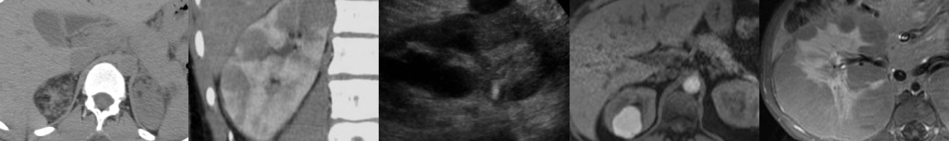
Ultrasound of Ovarian Dermoid Cyst
In this radiology lecture, we discuss the ultrasound appearance of ovarian dermoid cyst, including the rarely seen but highly specific “floating sphere” sign!
Key points include
- AKA mature cystic teratoma.
- Most common ovarian neoplasm.
- Benign, mean age 30.
- 10% bilateral.
- Mature tissue from ≥2 embryonic germ cell layers: Sebaceous material, hair follicles, skin derivatives, fat, muscle, bone, and other tissues lined by squamous epithelium.
- Specificity of US diagnosis 94-100%.
- MRI for changing morphology on f/u and for postmenopausal patients.
- Ultrasound findings: Floating echogenic spherical structures = “Floating sphere” sign (uncommon but pathognomonic), hyperechoic component with acoustic shadowing (Rokitanksy nodule), hyperechoic lines and dots, fat-fluid levels, diffuse or regional bright echoes.
- Most common complication: Ovarian torsion.
- Rare complications: Rupture, infection, malignant transformation, hormone secretion, anti-NMDA receptor encephalitis.
To learn more about the Samsung RS85 Prestige ultrasound system, please visit: https://www.bostonimaging.com/rs85-prestige-ultrasound-system-4
Click the YouTube Community tab or follow on social media for bonus teaching material posted throughout the week!
Instagram: https://www.instagram.com/radiologistHQ/
Facebook: https://www.facebook.com/radiologistHeadQuarters/
Twitter: https://twitter.com/radiologistHQ
Podcast: Play in new window | Download
Subscribe: Email