Case of the Week: Wandering Spleen (CT)
In this radiology lecture, we discuss the CT appearance of wandering spleen!
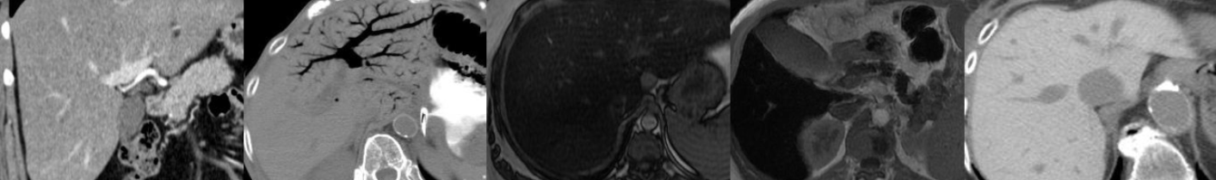
Key points include:
- Extremely rare, usually between 20-40 years of age, more common in females.
- Splenic mobility due to congenital or acquired abnormality of the normal peritoneal attachments/suspensory ligaments.
- Splenic migration to lower abdomen/pelvis, may develop long vascular pedicle.
- Twisting of pedicle can lead to splenic ischemia and infarction if not promptly treated.
- Variable clinical presentation, patients often become symptomatic if torsion of pedicle occurs: Intermittent colicky pain, vague abdominal discomfort, abdominal mass, acute abdomen.
- Treatment: Surgical detorsion and fixation of spleen (splenopexy), splenectomy may be required in setting of infarction.
Click the YouTube Community tab or follow on social media for bonus teaching material posted throughout the week!
Instagram: https://www.instagram.com/radiologistHQ/
Facebook: https://www.facebook.com/radiologistHeadQuarters/
Twitter: https://twitter.com/radiologistHQ
Podcast: Play in new window | Download
Subscribe: Email