Ultrasound of Epididymitis & Orchitis

In this radiology lecture, we review the ultrasound appearance of acute epididymitis and orchitis!

Key teaching points include:

  • Epididymitis = Inflammation of epididymis. Usually bacterial, most commonly due to retrograde ascent from bladder or prostate.
  • Causative infectious agent varies based on age: Adults younger than 35: Neisseria gonorrhoeae, Chlamydia trachomatis (STDs). Adults older than 35: E. coli & other coliform bacteria.
  • Non-infectious causes of epididymitis: Trauma, repetitive activities such as sports (most common causes in males prior to sexual maturity), torsed appendix testis or appendix epididymis, vasculitis, and medications (amiodarone).
  • Presentation: Gradual onset of scrotal pain, swelling & urinary symptoms. Must exclude testicular torsion (usually more acute onset of pain).
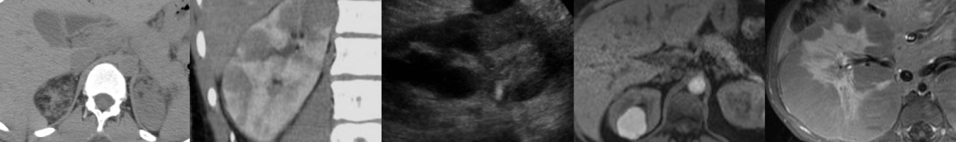
  • Epididymitis US findings: Epididymal enlargement, hyperemia, hypoechogenicity. Hyperemia usually precedes grey scale changes. Infection usually spreads from tail to body and head.
  • 20-30% of epididymitis cases have associated orchitis: Scrotal infection typically starts with epididymis then spreads to testis, scrotal sac, or scrotal wall.
  • Orchitis is less common than and usually secondary to epididymitis. Isolated orchitis uncommon, usually viral (mumps).
  • Orchitis US findings: Testicular enlargement, hyperemia and hypoechogenicity.
  • Complications: Scrotal wall inflammation, complicated hydrocele, pyocele (purulent fluid collection with mass effect), abscess (epididymal, testicular, scrotal wall), testicular ischemia and infarct due to obstructed venous outflow (decreased color Doppler testicular blood flow or reversed testicular diastolic arterial flow).

To learn more about the Samsung RS85 Prestige ultrasound system, please visit: https://www.bostonimaging.com/rs85-prestige-ultrasound-system-4

Click the YouTube Community tab or follow on social media for bonus teaching material posted throughout the week!

Spotify: https://bit.ly/spotify-rhq
Instagram: https://www.instagram.com/radquarters/
Facebook: https://www.facebook.com/radquarters/
Twitter: https://twitter.com/radquarters
Reddit: https://www.reddit.com/user/radiologistHQ/

Ultrasound of Torsion of the Appendix Testis

In this radiology lecture, we review the ultrasound appearance of torsion of the appendix testis and appendix epididymis!

Key teaching points include:

  • Appendix testis is a vestigial appendage usually located between upper pole of testis and head of epididymis.
  • AKA hydatid of Morgagni, the appendix testis is commonly present as a normal finding.
  • Appendix epididymis typically arises from epididymal head.
  • Both scrotal appendages are often pedunculated which increases risk of torsion.
  • Torsion occurs when appendage twists, occluding blood supply.
  • Torsion of the appendix testis is one of most common causes of acute scrotal pain in prepubertal children.
  • Peak age 7-12 years old, but can occur at any age.
  • Normal appendix testis: Oval-shaped, less than 6 mm in size, homogeneously isoechoic to epididymis, and demonstrates little to no blood flow on color Doppler.
  • Torsed appendix testis: 6 mm or larger in size, variable echogenicity, hypoechoic before 24 hours, hyperechoic or heterogeneous after 24 hours.
  • In setting of appendix torsion, hyperemia of surrounding structures with hydrocele and scrotal wall thickening often present.
  • Torsed appendage can detach and become free floating in scrotum.
  • Patients may present with pain localized to upper pole of testis or epididymis.
  • Physical examination may yield the “blue dot” sign: Small, palpable nodule at superior aspect of testis with bluish discoloration of overlying skin due to ischemic appendix.
  • Cremasteric reflex typically intact, and testicle not high riding (unlike testicular torsion).
  • Hyperemia of surrounding structures can be difficult to differentiate from bacterial epididymitis.
  • However, in children, epididymitis usually secondary to inflammation from direct trauma, torsion of a scrotal appendage, or urine reflux into epididymis. Urine dipstick/urinalysis helpful to differentiate from infection.
  • Treatment: Pain management with analgesics, ice, rest. If not recognized, may be treated unnecessarily with antibiotics. Scrotal exploration may be necessary if testicular torsion cannot be excluded.

References:
Baldisserotto M, Ketzer de Souza JC, Pertence AP, Dora MD. Color Doppler sonography of normal and torsed testicular appendages in children. AJR 2005; 184:1287–1292

To learn more about the Samsung RS85 Prestige ultrasound system, please visit: https://www.bostonimaging.com/rs85-prestige-ultrasound-system-4

Click the YouTube Community tab or follow on social media for bonus teaching material posted throughout the week!

Website: https://radiologisthq.com/
Spotify Video Podcast: https://bit.ly/spotify-rhq
Instagram: https://www.instagram.com/radiologistHQ/
Facebook: https://www.facebook.com/radiologistHeadQuarters/
Twitter: https://twitter.com/radiologistHQ
Reddit: https://www.reddit.com/user/radiologistHQ/

Ultrasound of Varicocele

In this radiology lecture, we review the ultrasound appearance of scrotal varicocele with three unique cases.

Key teaching points include:

  • Varicocele is abnormal dilatation of pampiniform venous plexus = Peritesticular veins.
  • Seen in up to 15% of adult and adolescent males.
  • Caused by incompetent or absent testicular vein valves.
  • Upper limit of normal for scrotal vein caliber = 2 mm, varicocele when greater than 2-3 mm.
  • Flow in varicocele usually too slow to detect with color Doppler and is typically better seen with Valsalva or with standing position.
  • 85% left sided, 15% bilateral: Left testicular vein drains into left renal vein at 90-degree angle, and superior mesenteric artery compresses left renal vein = Increased pressure and venous backflow. Right vein drains into IVC at acute angle.
  • Symptoms: Scrotal mass, pain, infertility/subfertility.
  • Low grade: Reflux only seen with Valsalva, inguinal canal/supratesticular location, vessels enlarged only in standing position.
  • High grade: Reflux seen at rest, infratesticular location, vessels enlarged in supine position.
  • Solitary right varicocele raises concern for compression of the right testicular vein from a retroperitoneal mass.
  • Ultrasound of upper abdomen should be considered when an isolated right-sided varicocele or asymmetrically large right-sided varicocele found.
  • However, most patients typically present with additional signs and symptoms of malignancy: “No patient in our cohort was found to have an unsuspected malignancy for which isolated right-sided varicocele was the only presenting sign.”*

*Gleason A, Bishop K, Xi Y et al. Isolated Right-Sided Varicocele: Is Further Workup Necessary? AJR 2019; 212:802-807

To learn more about the Samsung RS85 Prestige ultrasound system, please visit: https://www.bostonimaging.com/rs85-prestige-ultrasound-system-4

Click the YouTube Community tab or follow on social media for bonus teaching material posted throughout the week!

Instagram: https://www.instagram.com/radiologistHQ/
Facebook: https://www.facebook.com/radiologistHeadQuarters/
Twitter: https://twitter.com/radiologistHQ
Reddit: https://www.reddit.com/user/radiologistHQ/

Ultrasound of Testicular Torsion

In this radiology lecture, we review the ultrasound appearance of testicular torsion through three unique cases.

Key teaching points include:

  • Torsion occurs when spermatic cord twists and cuts off blood supply to the testis.
  • Bell-clapper deformity most common etiology: Abnormally high attachment of tunica vaginalis allowing spermatic cord rotation and testicular torsion (intravaginal).
  • Torsion has a bimodal distribution: First year of life (extravaginal), adolescents/young adults (intravaginal).
  • “Whirlpool” sign: Eddy swirl of coiled spermatic cord superior to testis, highly specific but less commonly seen than redundant spermatic cord.
  • Redundant spermatic cord AKA boggy pseudomass, torsion knot, epididymal-cord complex and should be avascular or only minimally vascular (unlike paratesticular neoplasm or acute epididymitis).
  • Testicles normally lie vertically, but horizontal or oblique (diagonal) lie suspicious for torsion.
  • Testicular enlargement, reactive hydrocele and scrotal skin thickening are secondary findings of torsion.
  • Marked testicular heterogeneity = Late torsion and nonviability/necrosis, more likely after 24 hours of symptoms.
  • Treatment: Detorsion and orchiopexy if salvageable, orchiectomy if not.

Reference: Bandarkar AN, Blask AR. Testicular torsion with preserved flow: Key sonographic features and value-added approach to diagnosis. Pediatric Radiology (2018) 48:735–744.

To learn more about the Samsung RS85 Prestige ultrasound system, please visit: https://www.bostonimaging.com/rs85-prestige-ultrasound-system-4

Click the YouTube Community tab or follow on social media for bonus teaching material posted throughout the week!

Instagram: https://www.instagram.com/radiologistHQ/
Facebook: https://www.facebook.com/radiologistHeadQuarters/
Twitter: https://twitter.com/radiologistHQ

Ultrasound & CT of Renal Oncocytoma

In this radiology lecture, the ultrasound and CT appearance of renal oncocytoma is revealed.

Key teaching points include:

  • Oncocytomas are benign, solid tumors.
  • 13% patients have multiple oncocytomas, and 1/3rd have concurrent renal cell carcinoma.
  • Central stellate nonenhancing scar only seen in 1/3rd of cases, and more commonly in larger tumors.
  • Spoke-wheel angiographic pattern may be present, best visualized on ultrasound using microvascular flow, AKA superb microvascular imaging.
  • Often not possible to differentiate from renal cell carcinoma (RCC) with imaging.
  • Both oncocytoma and RCC can have central scar and/or spoke-wheel angiographic pattern.
  • Kim et al.* found segmental enhancement inversion (corticomedullary phase/early excretory phase) characteristic for oncocytoma, but subsequent studies have shown inconsistent results.
  • When evaluating a renal mass, if only have postcontrast and 15-minute delayed phase images, mass deenhancement of 15 HU or more suggests a solid mass, whereas no change is more consistent with a hyperattenuating cyst.**

*Kim JI, Cho JY, Moon KC, et al. Segmental enhancement inversion at biphasic multidetector CT: Characteristic finding of small renal oncocytoma. Radiology 2009;252(2):441–448.

**Macari M, Bosniak MA. Delayed CT to evaluate renal masses incidentally discovered at contrast-enhanced CT: Demonstration of vascularity with deenhancement. Radiology 1999;213:674-680.

To learn more about the Samsung RS85 Prestige ultrasound system, please visit: https://www.bostonimaging.com/rs85-prestige-ultrasound-system-4

Click the YouTube Community tab or follow on social media for bonus teaching material posted throughout the week!

Instagram: https://www.instagram.com/radiologistHQ/
Facebook: https://www.facebook.com/radiologistHeadQuarters/
Twitter: https://twitter.com/radiologistHQ

Case of the Week: Ovarian Mucinous Cystadenocarcinoma (Ultrasound & MRI)

In this radiology lecture, we reveal the imaging appearance of mucinous cystadenocarcinoma of the ovary and explain differentiating features from serous cystadenocarcinoma.

Key points include:

  • A rare type of malignant ovarian epithelial tumor.
  • Often large at presentation, may be enormous.
  • Almost always multilocular.
  • Mucinous, proteinaceous and hemorrhagic material within loculi.
  • US: Scattered low-level echoes.
  • MRI: “Stained glass” appearance = Variable T1/T2 signal. Thick mucin = T1/T2 hyperintense.
  • Irregular, thick septations and solid components with internal vascularity and enhancement allow differentiation from mucinous cystadenoma.

Click the YouTube Community tab or follow on social media for bonus teaching material posted throughout the week!

Instagram: https://www.instagram.com/radiologistHQ/
Facebook: https://www.facebook.com/radiologistHeadQuarters/
Twitter: https://twitter.com/radiologistHQ

Case of the Week: Septate Uterus (MRI)

In this radiology lecture, we discuss the MRI appearance of septate uterus, and explain how to differentiate from other uterine anomalies.

Key points include:

  • Most common müllerian duct anomaly (55%): Septal reabsorption abnormality.
  • Ultrasound and MRI provide assessment of external uterine contour and presence of renal anomalies.
  • Hysterosalpingogram of limited value, cannot reliably differentiate between subtypes.
  • On MRI, uterine fundus is typically convex or minimally indented: Fundal cleft less than 1 cm.
  • Midline septum of variable length, may be muscular or fibrous.
  • Important to differentiate type of septum as may alter surgical approach.
  • Compared to bicornuate uterus, higher incidence of reproductive complications (miscarriage).
  • Treatment: Resection of septum if recurrent fetal loss.

Click the YouTube Community tab or follow on social media for bonus teaching material posted throughout the week!

Instagram: https://www.instagram.com/radiologistHQ/
Facebook: https://www.facebook.com/radiologistHeadQuarters/
Twitter: https://twitter.com/radiologistHQ

Ultrasound of Ovarian Dermoid Cyst

In this radiology lecture, we discuss the ultrasound appearance of ovarian dermoid cyst, including the rarely seen but highly specific “floating sphere” sign!

Key points include

  • AKA mature cystic teratoma.
  • Most common ovarian neoplasm.
  • Benign, mean age 30.
  • 10% bilateral.
  • Mature tissue from ≥2 embryonic germ cell layers: Sebaceous material, hair follicles, skin derivatives, fat, muscle, bone, and other tissues lined by squamous epithelium.
  • Specificity of US diagnosis 94-100%.
  • MRI for changing morphology on f/u and for postmenopausal patients.
  • Ultrasound findings: Floating echogenic spherical structures = “Floating sphere” sign (uncommon but pathognomonic), hyperechoic component with acoustic shadowing (Rokitanksy nodule), hyperechoic lines and dots, fat-fluid levels, diffuse or regional bright echoes.
  • Most common complication: Ovarian torsion.
  • Rare complications: Rupture, infection, malignant transformation, hormone secretion, anti-NMDA receptor encephalitis.

To learn more about the Samsung RS85 Prestige ultrasound system, please visit: https://www.bostonimaging.com/rs85-prestige-ultrasound-system-4

Click the YouTube Community tab or follow on social media for bonus teaching material posted throughout the week!

Instagram: https://www.instagram.com/radiologistHQ/
Facebook: https://www.facebook.com/radiologistHeadQuarters/
Twitter: https://twitter.com/radiologistHQ

Case of the Week: Retroperitoneal Fibrosis (Ultrasound & CT)

In this radiology lecture, we discuss the ultrasound and CT appearance of retroperitoneal fibrosis.

Key points include:

  • Most cases (70%) are idiopathic = Ormond disease.
  • Nonspecific symptoms depending on involved structures: Malaise, weight loss, low-grade fever.
  • Ureteral entrapment: Obstructive uropathy or renal failure, may see medial deviation of middle third of ureters with hydronephrosis.
  • Venous entrapment: Lower extremity edema, deep venous thrombosis.
  • CT: Soft tissue mass anterolateral to aorta with posterior sparing.
  • DDx: Retroperitoneal Lymphoma will Lift the aorta.
  • MRI: Low T1/T2 signal when inactive, T2 bright with early enhancement when active inflammation.
  • PET/CT: Avid when metabolically active, may aid in identifying appropriate biopsy sites.

Click the YouTube Community tab or follow on social media for bonus teaching material posted throughout the week!

Instagram: https://www.instagram.com/radiologistHQ/
Facebook: https://www.facebook.com/radiologistHeadQuarters/
Twitter: https://twitter.com/radiologistHQ

Case of the Week: Medullary Sponge Kidney (Ultrasound & CT)

Join me in this radiology lecture revealing the ultrasound and CT appearance of medullary sponge kidney (MSK).

Key points include:

  • MSK is a developmental ectasia with cystic dilatation of the collecting tubules in the pyramids leading to medullary nephrocalcinosis.
  • DDx medullary nephrocalcinosis: Hyperparathyroidism (most common cause in adults), renal tubular acidosis (type 1), MSK, hypervitaminosis D, other causes of hypercalcemia, sarcoidosis.
  • MSK associations: Beckwith-Wiedemann syndrome, congenital hemihypertrophy, Caroli disease, Ehlers-Danlos syndrome.
  • US: Echogenic medullary pyramids.
  • CT: Renal calculi, striated nephrogram, excretory phase “paintbrush” appearance or “growing calculus” sign.
  • Often asymptomatic but may present due to renal stones.

Click the YouTube Community tab or follow on social media for bonus teaching material posted throughout the week!

Instagram: https://www.instagram.com/radiologistHQ/
Facebook: https://www.facebook.com/radiologistHeadQuarters/
Twitter: https://twitter.com/radiologistHQ

1 2 3