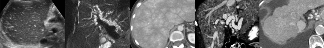
Ultrasound of Testicular Torsion
In this radiology lecture, we review the ultrasound appearance of testicular torsion through three unique cases.
Key teaching points include:
- Torsion occurs when spermatic cord twists and cuts off blood supply to the testis.
- Bell-clapper deformity most common etiology: Abnormally high attachment of tunica vaginalis allowing spermatic cord rotation and testicular torsion (intravaginal).
- Torsion has a bimodal distribution: First year of life (extravaginal), adolescents/young adults (intravaginal).
- “Whirlpool” sign: Eddy swirl of coiled spermatic cord superior to testis, highly specific but less commonly seen than redundant spermatic cord.
- Redundant spermatic cord AKA boggy pseudomass, torsion knot, epididymal-cord complex and should be avascular or only minimally vascular (unlike paratesticular neoplasm or acute epididymitis).
- Testicles normally lie vertically, but horizontal or oblique (diagonal) lie suspicious for torsion.
- Testicular enlargement, reactive hydrocele and scrotal skin thickening are secondary findings of torsion.
- Marked testicular heterogeneity = Late torsion and nonviability/necrosis, more likely after 24 hours of symptoms.
- Treatment: Detorsion and orchiopexy if salvageable, orchiectomy if not.
Reference: Bandarkar AN, Blask AR. Testicular torsion with preserved flow: Key sonographic features and value-added approach to diagnosis. Pediatric Radiology (2018) 48:735–744.
To learn more about the Samsung RS85 Prestige ultrasound system, please visit: https://www.bostonimaging.com/rs85-prestige-ultrasound-system-4
Click the YouTube Community tab or follow on social media for bonus teaching material posted throughout the week!
Instagram: https://www.instagram.com/radiologistHQ/
Facebook: https://www.facebook.com/radiologistHeadQuarters/
Twitter: https://twitter.com/radiologistHQ
Podcast: Play in new window | Download
Subscribe: Email