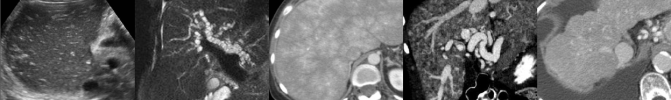
Ultrasound & CT of Renal Oncocytoma
In this radiology lecture, the ultrasound and CT appearance of renal oncocytoma is revealed.
Key teaching points include:
- Oncocytomas are benign, solid tumors.
- 13% patients have multiple oncocytomas, and 1/3rd have concurrent renal cell carcinoma.
- Central stellate nonenhancing scar only seen in 1/3rd of cases, and more commonly in larger tumors.
- Spoke-wheel angiographic pattern may be present, best visualized on ultrasound using microvascular flow, AKA superb microvascular imaging.
- Often not possible to differentiate from renal cell carcinoma (RCC) with imaging.
- Both oncocytoma and RCC can have central scar and/or spoke-wheel angiographic pattern.
- Kim et al.* found segmental enhancement inversion (corticomedullary phase/early excretory phase) characteristic for oncocytoma, but subsequent studies have shown inconsistent results.
- When evaluating a renal mass, if only have postcontrast and 15-minute delayed phase images, mass deenhancement of 15 HU or more suggests a solid mass, whereas no change is more consistent with a hyperattenuating cyst.**
*Kim JI, Cho JY, Moon KC, et al. Segmental enhancement inversion at biphasic multidetector CT: Characteristic finding of small renal oncocytoma. Radiology 2009;252(2):441–448.
**Macari M, Bosniak MA. Delayed CT to evaluate renal masses incidentally discovered at contrast-enhanced CT: Demonstration of vascularity with deenhancement. Radiology 1999;213:674-680.
To learn more about the Samsung RS85 Prestige ultrasound system, please visit: https://www.bostonimaging.com/rs85-prestige-ultrasound-system-4
Click the YouTube Community tab or follow on social media for bonus teaching material posted throughout the week!
Instagram: https://www.instagram.com/radiologistHQ/
Facebook: https://www.facebook.com/radiologistHeadQuarters/
Twitter: https://twitter.com/radiologistHQ
Podcast: Play in new window | Download
Subscribe: Email