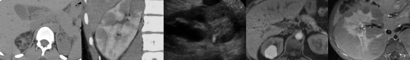
Case of the Week: Perihilar Cholangiocarcinoma/Klatskin Tumor (CT & MRI)

In this radiology lecture, we discuss the CT and MRI appearance of perihilar cholangiocarcinoma.

Key points include:

  • Perihilar cholangiocarcinoma (AKA Klatskin tumor) occurs at bifurcation of the hepatic duct.
  • Cholangiocarcinoma (CC) is a primary malignant tumor of bile duct epithelium, usually adenocarcinoma.
  • CC is the most common primary hepatic malignancy after hepatocellular carcinoma (HCC), and most are extrahepatic (as opposed to intrahepatic).
  • Appearance of CC is based on growth pattern: Mass-forming, periductal infiltrating, and intraductal growing.
  • Risk factors: Parasite infection, choledochal cyst, primary sclerosing cholangitis, recurrent pyogenic cholangitis, and inflammatory bowel disease (ulcerative colitis).
  • Patients are usually 65 or older.
  • On CT and MRI, perihilar CC appears as a biliary stricture with shouldering/abrupt tapering.
  • If a mass is visible, will typically have rimlike enhancement with gradual centripetal enhancement on delayed images, be T2 bright (but not as homogeneous or as bright as hemangioma), and may have a targetlike appearance on DWI (favors CC over HCC).

Click the YouTube Community tab or follow on social media for bonus teaching material posted throughout the week!

Instagram: https://www.instagram.com/radiologistHQ/
Facebook: https://www.facebook.com/radiologistHeadQuarters/
Twitter: https://twitter.com/radiologistHQ


Case of the Week: Medullary Sponge Kidney (Ultrasound & CT)

Join me in this radiology lecture revealing the ultrasound and CT appearance of medullary sponge kidney (MSK).

Key points include:

  • MSK is a developmental ectasia with cystic dilatation of the collecting tubules in the pyramids leading to medullary nephrocalcinosis.
  • DDx medullary nephrocalcinosis: Hyperparathyroidism (most common cause in adults), renal tubular acidosis (type 1), MSK, hypervitaminosis D, other causes of hypercalcemia, sarcoidosis.
  • MSK associations: Beckwith-Wiedemann syndrome, congenital hemihypertrophy, Caroli disease, Ehlers-Danlos syndrome.
  • US: Echogenic medullary pyramids.
  • CT: Renal calculi, striated nephrogram, excretory phase “paintbrush” appearance or “growing calculus” sign.
  • Often asymptomatic but may present due to renal stones.

Click the YouTube Community tab or follow on social media for bonus teaching material posted throughout the week!

Instagram: https://www.instagram.com/radiologistHQ/
Facebook: https://www.facebook.com/radiologistHeadQuarters/
Twitter: https://twitter.com/radiologistHQ

Case of the Week: Amebic Liver Abscess (Ultrasound & CT)

In this radiology lecture, we discuss the ultrasound and CT appearance of amebic liver abscess.

Key points include:

  • Entamoeba histolytica infection.
  • Endemic in Africa, Southeast Asia, and Central & South America.
  • More common in males.
  • Presents as right upper quadrant pain, fever and hepatomegaly.
  • Both amebic and pyogenic (bacterial) abscesses can have a layered wall with the “double target” or “double rim” sign.
  • Amebic more likely to be unilocular (septations present in 30%) without “cluster” sign typical of multiloculated pyogenic abscess.
  • Amebic more likely solitary, pyogenic more likely multiple.
  • Can be treated medically (metronidazole), but if diagnosis uncertain, if there is failed response to medical therapy, or if large abscess at risk for rupture = aspiration.

Bächler P, Baladron MJ, Menias C, et al. Multimodality Imaging of Liver Infections: Differential Diagnosis and Potential Pitfalls. RadioGraphics 2016 36:4, 1001-1023.

Click the YouTube Community tab or follow on social media for bonus teaching material posted throughout the week!

Instagram: https://www.instagram.com/radiologistHQ/
Facebook: https://www.facebook.com/radiologistHeadQuarters/
Twitter: https://twitter.com/radiologistHQ

Case of the Week: Pulmonary Infarction (X-ray & CT)

In this radiology lecture, we discuss the chest x-ray and CT appearance of pulmonary infarction in the setting of acute pulmonary embolism.

Key points include:

  • Uncommon complication of pulmonary embolism.
  • Most common in right lung.
  • Risk of infarction increases with large clot burden.
  • Typically wedge-shaped, peripheral consolidation with no air bronchograms (Hampton hump).
  • However, may not be wedge-shaped, and not all wedge-shaped opacities will be infarcts in the setting of pulmonary embolism.
  • “Bubbly” consolidation containing rounded, central lucencies: Most specific finding of infarct* and represents a combination of infarcted, necrotic lung and adjacent viable, aerated lung.
  • “Vessel” sign: Enlarged vessel leading to apex of a wedge-shaped opacity. Vessel is dilated due to the presence of intraluminal thrombus or distal obstruction.

*Revel MP, Triki R, Chatellier G, et al. Is it possible to recognize pulmonary infarction on multisection CT images? Radiology. 2007;244(3):875-882.

Click the YouTube Community tab or follow on social media for bonus teaching material posted throughout the week!

Instagram: https://www.instagram.com/radiologistHQ/
Facebook: https://www.facebook.com/radiologistHeadQuarters/
Twitter: https://twitter.com/radiologistHQ

Case of the Week: Ruptured Ectopic Pregnancy (Ultrasound)

In this radiology lecture, we discuss the ultrasound appearance of ruptured ectopic pregnancy.

Key points include:

  • Most ectopic pregnancies occur in the fallopian tube: Ampulla most common, followed by isthmus and fimbria.
  • Risk factors: Prior ectopic pregnancy, prior surgery (fallopian tube), pelvic inflammatory disease, endometriosis, IVF.
  • “A single measurement of hCG, regardless of its level, does not reliably distinguish between ectopic and intrauterine pregnancy (viable or nonviable).”*
  • Levels of hCG in ectopic pregnancies are highly variable.
  • Tubal rupture main complication, occurs in up to 20%.
  • Free fluid in pelvis alone nonspecific, but echogenic fluid in Morison pouch (subhepatic space) and cul-de-sac raises concern for rupture.
  • Rupture is a relative contraindication to methotrexate (medical) therapy.

*Doubilet PM, Benson CB, Bourne T, et al. Diagnostic criteria for nonviable pregnancy early in the first trimester. N Engl J Med 2013;369:1443-51.

Click the YouTube Community tab or follow on social media for bonus teaching material posted throughout the week!

Instagram: https://www.instagram.com/radiologistHQ/
Facebook: https://www.facebook.com/radiologistHeadQuarters/
Twitter: https://twitter.com/radiologistHQ

Case of the Week: Gallstone Ileus (X-ray & CT)

In this radiology lecture, we discuss the appearance of gallstone ileus on x-ray and CT.

Key points include:

  • Gallstone ileus is a rare complication of chronic cholecystitis.
  • Actually not an ileus, but a small bowel obstruction.
  • Gallstone migrates through a fistula between gallbladder and small bowel (usually duodenum) and becomes impacted in the terminal ileum.
  • Stone can also impact in the proximal ileum, jejunum, even in the duodenum/distal stomach causing gastric outlet obstruction (Bouveret syndrome).
  • Rigler triad on abdominal x-ray: Small bowel obstruction, pneumobilia and gallstone in the right iliac fossa.
  • Usually affects the elderly and treated surgically.

Click the YouTube Community tab or follow on social media for bonus teaching material posted throughout the week!

Instagram: https://www.instagram.com/radiologistHQ/
Facebook: https://www.facebook.com/radiologistHeadQuarters/
Twitter: https://twitter.com/radiologistHQ

Case of the Week: Testicular Epidermoid Cyst (Ultrasound)

In this radiology lecture, we discuss the ultrasound appearance of testicular epidermoid cyst.

Key points include:

  • Testicular epidermoid cyst is a rare, benign, intratesticular neoplasm.
  • Most common in 2nd-4th decades, typically presents as a painless mass.
  • Lamellated, onion-like, bull’s-eye appearance: Alternating hyperechoic and hypoechoic concentric rings.
  • Appearance secondary to cyst filled with layers of keratin and lined with keratinizing squamous epithelium.
  • Non-vascular and sharply marginated.
  • Nonenhancing on MRI.
  • Important to recognize preoperatively because may be treated with conservative surgery.
  • Management somewhat controversial as originally diagnosed with orchiectomy.
  • Increasingly treated with enucleation if frozen sections of mass are consistent and tumor markers are negative.

Click the YouTube Community tab or follow on social media for bonus teaching material posted throughout the week!

Instagram: https://www.instagram.com/radiologistHQ/
Facebook: https://www.facebook.com/radiologistHeadQuarters/
Twitter: https://twitter.com/radiologistHQ

Case of the Week: Necrotizing Pancreatitis (CT & MRI)

In this radiology lecture, we discuss the imaging appearance of necrotizing pancreatitis on both CT and MRI.

Key points include:

  • According to the revised Atlanta classification, there are two types of acute pancreatitis: Interstitial edematous pancreatitis (IEP) and necrotizing pancreatitis (NP).
  • For IEP, fluid collection in first 4 weeks = acute peripancreatic fluid collection, after 4 weeks = pseudocyst.
  • For NP, fluid collection in first 4 weeks = acute necrotic collection, after 4 weeks = walled-off necrosis.
  • Non-enhancing hypoattenuating areas = necrotizing pancreatitis.
  • Gas suspicious for infection/emphysematous pancreatitis.
  • Vascular complications are important to identify.
  • Venous thrombosis: splenic, portal, and mesenteric veins.
  • Pseudoaneurysms: Splenic and gastroduodenal artery.

Click the YouTube Community tab or follow on social media for bonus teaching material posted throughout the week!

Instagram: https://www.instagram.com/radiologistHQ/
Facebook: https://www.facebook.com/radiologistHeadQuarters/
Twitter: https://twitter.com/radiologistHQ

Case of the Week: Duplicated Collecting System (VCUG & Ultrasound)

In this radiology lecture, we discuss the imaging appearance of duplicated collecting system and ureterocele, with attention to US and VCUG.

Key points include:

  • Weigert-Meyer rule: Remember the mnemonic “DUMI.”
  • With duplex kidneys and complete ureteral Duplication, ureter draining Upper pole inserts ectopically into bladder Medially and Inferiorly to ureter draining lower pole.
  • Lower pole moiety inserts orthotopically.
  • Upper pole moiety often ends as an ectopic ureterocele.
  • Upper pole moiety tends to obstruct, and lower pole moiety is prone to reflux.
  • Obstructed upper pole moiety causes mass effect with resultant inferior displacement of the lower pole moiety and the “drooping lily” sign.

Click the YouTube Community tab or follow on social media for bonus teaching material posted throughout the week!

Instagram: https://www.instagram.com/radiologistHQ/
Facebook: https://www.facebook.com/radiologistHeadQuarters/
Twitter: https://twitter.com/radiologistHQ

Case of the Week: Colonic Lymphoma (CT & PET)

In this radiology lecture, we discuss the imaging appearance of large bowel lymphoma.

Key points include:

  • Often isodense to skeletal muscle.
  • May have aneurysmal dilatation of involved bowel.
  • Less likely obstructive and longer segment involvement compared to colonic adenocarcinoma.
  • Located near ileocecal valve.
  • GI lymphoma: Most common in stomach, followed by small bowel (ileum, jejunum, duodenum), least common site colorectal.
  • Splenomegaly and severe lymphadenopathy favor lymphoma but may not be present.

Click the YouTube Community tab or follow on social media for bonus teaching material posted throughout the week!

Instagram: https://www.instagram.com/radiologistHQ/
Facebook: https://www.facebook.com/radiologistHeadQuarters/
Twitter: https://twitter.com/radiologistHQ

1 3 4 5 6 7 8