Case of the Week: Colonic Lymphoma (CT & PET)
In this radiology lecture, we discuss the imaging appearance of large bowel lymphoma.
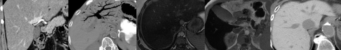
Key points include:
- Often isodense to skeletal muscle.
- May have aneurysmal dilatation of involved bowel.
- Less likely obstructive and longer segment involvement compared to colonic adenocarcinoma.
- Located near ileocecal valve.
- GI lymphoma: Most common in stomach, followed by small bowel (ileum, jejunum, duodenum), least common site colorectal.
- Splenomegaly and severe lymphadenopathy favor lymphoma but may not be present.
Click the YouTube Community tab or follow on social media for bonus teaching material posted throughout the week!
Instagram: https://www.instagram.com/radiologistHQ/
Facebook: https://www.facebook.com/radiologistHeadQuarters/
Twitter: https://twitter.com/radiologistHQ
Podcast: Play in new window | Download
Subscribe: Email