Case of the Week: Gallstone Ileus (X-ray & CT)
In this radiology lecture, we discuss the appearance of gallstone ileus on x-ray and CT.
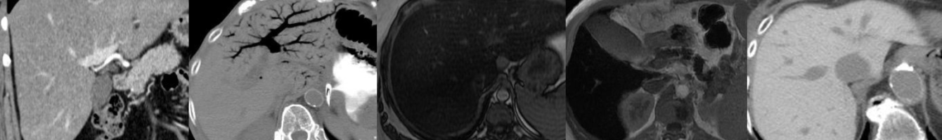
Key points include:
- Gallstone ileus is a rare complication of chronic cholecystitis.
- Actually not an ileus, but a small bowel obstruction.
- Gallstone migrates through a fistula between gallbladder and small bowel (usually duodenum) and becomes impacted in the terminal ileum.
- Stone can also impact in the proximal ileum, jejunum, even in the duodenum/distal stomach causing gastric outlet obstruction (Bouveret syndrome).
- Rigler triad on abdominal x-ray: Small bowel obstruction, pneumobilia and gallstone in the right iliac fossa.
- Usually affects the elderly and treated surgically.
Click the YouTube Community tab or follow on social media for bonus teaching material posted throughout the week!
Instagram: https://www.instagram.com/radiologistHQ/
Facebook: https://www.facebook.com/radiologistHeadQuarters/
Twitter: https://twitter.com/radiologistHQ
Podcast: Play in new window | Download
Subscribe: Email