Imaging of Pheochromocytoma

In this video lecture, we discuss the diagnosis and imaging appearance of pheochromocytoma on ultrasound, CT, MRI, and nuclear medicine studies.

Key points include:

  • Pheochromocytoma is a biochemical diagnosis.
  • Symptoms are variable, and patients may present with headache, palpitations, sweating, hypertension or be asymptomatic.
  • Most cases are sporadic with mean age of presentation in the 40s.
  • Hereditary pheochromocytoma can be seen in the setting of Von Hippel-Lindau disease, multiple endocrine neoplasia (MEN) type II, neurofibromatosis type I, and pheochromocytoma-paraganglioma syndromes associated with succinate dehydrogenase gene mutations.
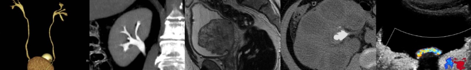
  • Pheochromocytoma is the “10%” tumor, as approximately 10% are bilateral, extra-adrenal, contain calcification, occur in children, or are malignant.
  • Most pheochromocytomas are solid, but they can also appear as mixed cystic and solid with varying degrees of complexity.
  • The ultrasound appearance is nonspecific, and tumors may contain echogenic hemorrhage or be hypervascular on Doppler imaging.
  • Any mass that enhances to a density greater than 110 HU on an arterial phase CT scan should raise suspicion for pheochromocytoma.
  • On CT, most pheochromocytomas will have noncontrast density greater than 10 HU and absolute washout less than 60%, findings similar to adrenal metastases and adrenocortical carcinoma.
  • However, pheochromocytoma can rarely have noncontrast density less than 10 HU and absolute washout greater than 60%, thus mimicking adrenal adenoma.
  • Studies have shown that pheochromocytoma is not a contraindication to modern nonionic IV contrast media.
  • On MRI, the “light bulb” T2 bright appearance classically associated with pheochromocytoma may or may not be present.
  • If intratumoral hemorrhage is present, pheochromocytomas may show T1 hyperintensity or susceptibility artifact on T1-weighted in-phase images depending on the age of blood.
  • Pheochromocytomas usually do not show signal dropout on T1-weighted opposed-phase images but rarely do and can mimic adrenal adenomas.
  • MIBG scans have nearly 100% specificity for pheochromocytoma and are useful to confirm clinically suspected pheochromocytoma and for detection of metastatic disease.
  • Extra-adrenal pheochromocytomas (paragangliomas) usually occur in abdomen, typically at the organ of Zuckerkandl or in the retroperitoneum.

Malignant Adrenal Masses

In this video lecture, we discuss the diagnosis and imaging appearance of malignant adrenal masses: adrenal metastases including collision tumor, adrenocortical carcinoma and adrenal lymphoma.

Key points include:

  • Adrenal metastases are the most common malignant lesion involving the adrenal gland.
  • Lung carcinoma is the most common primary malignancy to metastasize to the adrenal glands.
  • Adrenal metastases are often bilateral and greater than 3 cm in size.
  • When malignant adrenal lesions are compared to adenomas, SUV cutoff of 3.1 has a 99% negative predictive value.
  • Adrenal-to-liver SUV ratio cutoff value of 1.4 has a specificity of 100% in differentiating adrenal adenomas and metastases.
  • Collision tumors are two histologically distinct tumors that abut or are near each other in the adrenal gland, and PET/CT is the best way to characterize these lesions without biopsy.
  • An enlarging defect within adrenal signal dropout on T1-weighted opposed-phase GRE images is suspicious for a metastatic collision tumor abutting a lipid-rich adrenal adenoma.
  • Renal cell carcinoma metastases can be slow growing and occur many years after the initial tumor presentation.
  • Adrenocortical carcinoma has a bimodal age distribution, may be hormonally functioning and has a poor prognosis.
  • Adrenocortical carcinoma usually presents as a large (greater than 6 cm) mass with internal hemorrhage, necrosis and sometimes calcification.
  • Venous invasion is common with adrenocortical carcinoma.
  • Adrenal lymphoma will be round or adreniform in shape and frequently shows restricted diffusion, a feature that can be helpful in differentiating from adrenal hyperplasia.
  • Diffuse large B-cell lymphoma is the most common type of adrenal lymphoma, and patients usually present with B-cell symptoms and/or adrenal insufficiency.
  • Adrenal lymphoma is usually bilateral and may invade the adjacent kidney(s).

Imaging of Pelvic Inflammatory Disease, Part I

In this video lecture, we discuss the ultrasound and computed tomography (CT) appearance of pelvic inflammatory disease (PID).

Topics include:

  • Early findings of PID, including haziness of pelvic fat, salpingitis, oophoritis, and endometritis.
  • Importance of indentifying dilated fallopian tubes by the characteristic “C” or “S” shape that they exhibit, as well as the presence of the “cogwheel” sign.
  • Differentiating tubo-ovarian abscess (TOA) from tubo-ovarian complex (TOC), and the associated treatment implications.
  • Emphazing the typical posterior, dependent position of pyosalpinges and TOAs, as well as the associated anterior displacement of the broad ligament/mesosalpinx.

References:

Imaging of Pelvic Inflammatory Disease, Part II

In the second part of this video lecture, we discuss the MRI appearance of pelvic inflammatory disease (PID), including tips on how to differentiate pyosalpinx from both hydrosalpinx and hematosalpinx.  The computed tomography (CT) and ultrasound appearance of associated complications of PID are also reviewed.

Topics include:

  • Using diffusion-weighted imaging to differentiate pyosalpinx/tubo-ovarian abscess (TOA) from hydrosalpinx.
  • Association between hematosalpinx and endometriosis.
  • Fitz-Hugh-Curtis perihepatitis syndrome and its CT appearance on both late hepatic arterial phase and portal venous phase imaging.
  • Imaging appearance of endometritis, including fluid-filled dilated endometrial cavity, subendometrial enhancement, gas, “dirty” shadowing and (most importantly) presence of clinical suspicion.
  • Ovarian vein thrombosis, with emphasis on identifying enhancing, thickened venous walls and perivenous inflammation as findings of thrombophlebitis.
  • TOA secondary to diverticulitis.
  • How oxidized regenerated cellulose can mimic pelvic abscess and can normally contain gas for at least a month after surgery, highlighting the importance of reviewing the details of the operative history.

References:

Introduction to Genitourinary Radiology, Part II

In this video lecture, we “leave no stone unturned” as we introduce both renal and bladder calculi. Also, the basics of renal mass evaluation with ultrasound, computed tomography (CT), and magnetic resonance imaging (MRI) are discussed.

Topics include:

  • The importance of Hounsfield unit density when describing renal calculi.
  • CT and ultrasound appearance of nephrouterolithiasis and renal obstruction.
  • Basic retroperitoneal fascial plane anatomy.
  • Techniques to optimize stone detection with ultrasound.
  • How to identify whether you are looking at a T1- or T2-weighted MRI sequence.
  • Differentiating simple and hyperdense renal cysts from neoplasm with CT and MRI.
  • The importance of MRI subtraction imaging.

Introduction to Genitourinary Radiology, Part III

In the conclusion of this introductory series, we discuss the basics of computed tomography (CT) urography, bladder and urethral diverticula, and scrotal pathology including varicocele, epididymo-orchitis, and testicular neoplasm.

Topics include:

  • CT urography post-processing techniques: maximum intensity projection (MIP) and 3D volume-rendered (VR) imaging.
  • Appearance of transitional cell carcinoma in the ureters and bladder on excretory phase CT mages.
  • Papillary necrosis.
  • Weigert-Meyer rule for duplicated collecting systems and tips to remember it.
  • Urethral diverticula appearance on magnetic resonance imaging (MRI) and voiding cystourography (VCUG).
  • Scrotal varicocele and the importance of recognizing the implications of right-sided versus left-sided.
  • Acute scrotal pathology: testicular torsion and epididymo-orchitis.
  • Differentiating seminomatous and non-seminomatous testicular neoplasms via ultrasound.

1 2 3