Case of the Week: Testicular Epidermoid Cyst (Ultrasound)

In this radiology lecture, we discuss the ultrasound appearance of testicular epidermoid cyst.

Key points include:

  • Testicular epidermoid cyst is a rare, benign, intratesticular neoplasm.
  • Most common in 2nd-4th decades, typically presents as a painless mass.
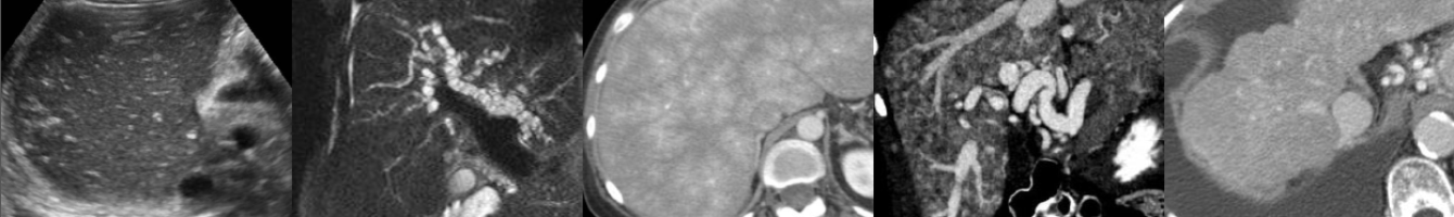
  • Lamellated, onion-like, bull’s-eye appearance: Alternating hyperechoic and hypoechoic concentric rings.
  • Appearance secondary to cyst filled with layers of keratin and lined with keratinizing squamous epithelium.
  • Non-vascular and sharply marginated.
  • Nonenhancing on MRI.
  • Important to recognize preoperatively because may be treated with conservative surgery.
  • Management somewhat controversial as originally diagnosed with orchiectomy.
  • Increasingly treated with enucleation if frozen sections of mass are consistent and tumor markers are negative.

Click the YouTube Community tab or follow on social media for bonus teaching material posted throughout the week!

Instagram: https://www.instagram.com/radiologistHQ/
Facebook: https://www.facebook.com/radiologistHeadQuarters/
Twitter: https://twitter.com/radiologistHQ

Case of the Week: Duplicated Collecting System (VCUG & Ultrasound)

In this radiology lecture, we discuss the imaging appearance of duplicated collecting system and ureterocele, with attention to US and VCUG.

Key points include:

  • Weigert-Meyer rule: Remember the mnemonic “DUMI.”
  • With duplex kidneys and complete ureteral Duplication, ureter draining Upper pole inserts ectopically into bladder Medially and Inferiorly to ureter draining lower pole.
  • Lower pole moiety inserts orthotopically.
  • Upper pole moiety often ends as an ectopic ureterocele.
  • Upper pole moiety tends to obstruct, and lower pole moiety is prone to reflux.
  • Obstructed upper pole moiety causes mass effect with resultant inferior displacement of the lower pole moiety and the “drooping lily” sign.

Click the YouTube Community tab or follow on social media for bonus teaching material posted throughout the week!

Instagram: https://www.instagram.com/radiologistHQ/
Facebook: https://www.facebook.com/radiologistHeadQuarters/
Twitter: https://twitter.com/radiologistHQ

Webinar: Ultrasound of Ovarian Cystic Disease

Check out my FREE webinar titled “Ultrasound of Ovarian Cystic Disease” on Wednesday, September 29, 2021 at 7-8 PM ET, hosted by the American Institute of Ultrasound in Medicine (AIUM). A Q&A session follows the presentation.

In case you missed the lecture or would like to watch it again, click here: https://learn.aium.org/products/ultrasound-of-cystic-ovarian-disease-neoplastic-non-neoplastic

Enjoy!

Webinar: Ultrasound of Genitourinary Infectious Disease

Check out my FREE webinar titled “Ultrasound of Genitourinary Infectious Disease” on Thursday, March 25, 2021 at 1-2 PM ET, hosted by the American Institute of Ultrasound in Medicine (AIUM). A Q&A session follows the presentation.

In case you missed the lecture or would like to watch it again, you can find it here: https://learn.aium.org/products/ultrasound-of-genitourinary-infectious-disease

Enjoy!

Non-masslike Adrenal Abnormalities

In this video lecture, we discuss the normal imaging appearance of the adrenal glands, as well as multiple common and rare non-masslike adrenal abnormalities on CT and MRI.

Key points include:

  • The adrenal glands normally have an inverted “V” or “Y” configuration.
  • A linear, disk-like adrenal gland or “pancake” adrenal gland indicates congenital malposition or absence of the ipsilateral kidney.
  • Adrenal hyperenhancement raises suspicion for developing shock and may signal a therapeutic window to act upon.
  • Adrenal calcifications can be caused by prior hemorrhage, infection, tumors and metabolic syndromes.
  • On MRI, T1-weighted in-phase GRE images can be helpful in identifying calcification due to dark blooming caused by susceptibility artifact.
  • Adrenal atrophy may indicate adrenal insufficiency.
  • Adrenal hyperplasia is identified when adrenal limbs measure greater than 10 mm in thickness and may or may not be hormonally functioning.
  • Nonhemorrhagic adrenal infarction appears as a nonenhancing, edematous adrenal gland with surrounding stranding and restricted diffusion.
  • Adrenal infarction can be seen in hypercoagulable states, antiphospholipid-antibody syndrome and pregnancy.

Imaging of Adrenal Adenomas & Incidentalomas

In this video lecture, we discuss the imaging appearance of lipid-rich and lipid-poor adrenal adenomas, explain the CT washout calculation, and review the choice of CT vs. MRI for the evaluation of adrenal nodules. Also, we compare the 2017 ACR (American College of Radiologists) Incidental Findings Committee recommendations to the AACE/AAES (American Association of Clinical Endocrinologists and American Association of Endocrine Surgeons) medical guidelines in the evaluation of adrenal incidentalomas.

Key points include:

  • 70% of adrenal adenomas will be lipid-rich with a low-density of 10 or less Hounsfield units (HU) on CT.
  • Adrenal nodules with greater than 60% enhancement washout and greater than 40% relative washout are consistent with adenomas.
  • Using chemical shift, MRI may be helpful in characterizing a mass which is indeterminate on non-contrast CT, particularly if the non-contrast density is less than 20-30 HU.
  • If the non-contrast density of an adrenal nodule is greater than 20-30 HU, MRI should be avoided, as washout CT is typically the test of choice.
  • While the AACE/AAES and ACR follow-up algorithms for adrenal nodules differ in many respects, they agree that surgical resection should be considered for adrenal masses measuring 4 cm or more in size, and that biochemical evaluation should at least be considered for most adrenal masses.

Adrenal Hemorrhage

In this video lecture, we discuss the imaging appearance of adrenal hemorrhage on CT, MRI and ultrasound. Causes of adrenal hemorrhage will also be reviewed. 

Key points include:

  • On CT scan, adrenal hemorrhage typically appears as a nonenhancing round or oval hyperdense mass with density of 50-90 HU. 
  • MRI is the most sensitive and specific modality for diagnosing adrenal hemorrhage. 
  • The “high signal intensity rim” rim sign seen on T1-weighted images is characteristic of subacute adrenal hemorrhage. 
  • Adrenal hemorrhage may appear solid or cystic on ultrasound depending on the age of hemorrhage.
  • Adrenal hemorrhage is more common in neonates than in children and adults and is the most common adrenal mass in infancy.
  • Trauma is the most common cause and is usually unilateral and right-sided. 
  • Atraumatic adrenal hemorrhage is usually unilateral.

Adrenal Cysts and Myelolipomas

In this video lecture, we discuss the imaging appearance of adrenal cysts and myelolipomas, as well as differential diagnostic considerations.

Key points include:

  • Adrenal cysts typically have density of simple fluid ranging from 20 to -10 Hounsfield units (HU).
  • Adrenal pseudocysts may be symptomatic, may have a complicated appearance and may have peripheral calcification.
  • The “claw” sign can be helpful in differentiating adrenal from renal masses.
  • Gastric diverticula can mimic adrenal cysts but can be differentiated by identifying communication of gastric diverticulum with adjacent stomach, as well as the presence of oral contrast and/or gas with the diverticulum.
  • Adrenal myelolipomas are benign neoplasms that have varying degrees of macroscopic fat and hematopoietic soft tissue elements.
  • Fat density within myelolipomas on CT ranges from -30 to -90 HU.
  • Up to 30% of myelolipomas contain small calcifications.
  • Unlike myelolipomas, malignant retroperitoneal sarcomas are usually ill-defined and displace or invade adjacent structures.
  • Because myelolipomas contain macroscopic fat, they show greater signal loss (darkening) on fat-saturated images compared to T1 opposed-phase chemical shift images.
  • Conversely, lipid-rich adrenal adenomas contain microscopic fat (also known as intracellular, intracytoplasmic, or intravoxel fat) and therefore show greater signal loss on T1 opposed-phase images than on fat-saturated images.
1 2 3