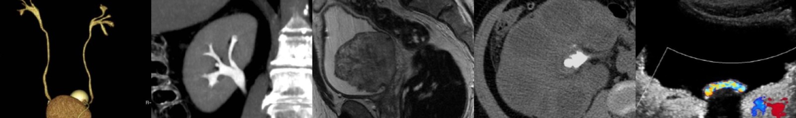
Ultrasound of Achilles Tendinosis and Tear
In this radiology lecture, we review the ultrasound appearance of Achilles tendinosis, partial thickness tears and full thickness tears through four unique cases.
Key teaching points include:
- Achilles tendon is strongest in body. Originates from soleus and gastrocnemius muscles, inserts onto posterior calcaneal tuberosity.
- Achilles tendon tears = Most common ankle tendon injury.
- Tendon enlargement greater than 1 cm in AP dimension = Abnormal.
- Tendinosis appears as fusiform hypoechoic swelling of tendon without fiber disruption with increased blood flow (use power Doppler or microvascular flow).
- Ultrasound highly sensitive and specific for partial and complete Achilles tears.
- Partial tear = Hypoechoic/anechoic cleft that disrupts tendon fibers.
- Full thickness tears = Usually 2-6 cm proximal to calcaneal insertion. Complete tendon fiber disruption and retraction. May see refractive shadowing at tendon stumps. Tendon gap may fill with mixed echogenicity fluid/hemorrhage or portion of adjacent fat pad.
- Plantaris tendon = Thin tendon at medial aspect of Achilles, may mimic intact Achilles tendon fibers (plantaris usually stays intact with Achilles tear).
- Dynamic imaging with passive ankle dorsiflexion and plantar flexion helps reveal tendon retraction at tear site.
- Achilles tendon surrounded by a paratenon as opposed to a true synovial tendon sheath.
- Paratendinitis = Hypoechoic swelling or anechoic fluid adjacent to tendon.
- Achilles tendon ossification can occur with prior tendon rupture, surgery, or repetitive microtrauma.
- Scar tissue in chronic tear can simulate tendon fibers (dynamic maneuvers helpful), and fibrous bridging may occur.
Click the YouTube Community tab or follow on social media for bonus teaching material posted throughout the week!
Instagram: https://www.instagram.com/radiologistHQ/
Facebook: https://www.facebook.com/radiologistHeadQuarters/
Twitter: https://twitter.com/radiologistHQ
Podcast: Play in new window | Download
Subscribe: Email