Ultrasound of Submandibular Sialolithiasis (Salivary Stones)
In this radiology lecture, we review the ultrasound and CT appearance of submandibular stone disease, together with floor of mouth anatomy!
Key teaching points include:
- Submandibular glands are paired salivary glands located inferior to body of mandible
- Submandibular glands are intermediate in size compared to the larger parotid and smaller sublingual glands, and they do not contain lymph nodes
- The submandibular duct (= Wharton’s duct) extends from gland hilum and travels superiorly to open at floor of mouth on either side of base of frenulum of tongue. The duct is not typically seen unless abnormally dilated
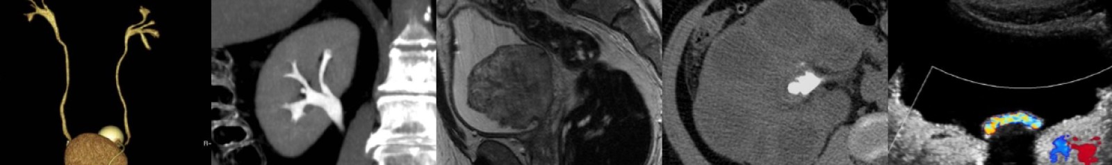
- On ultrasound, normal submandibular glands appear as encapsulated structures with homogeneous echotexture similar to the parotid. Fine linear echodensities may be present representing intraglandular ductules. Physiologic intravascular flow is typically evident. Superficial portion of the gland is almond-shaped, deep portion triangular
- Sialolithiasis = Salivary calculous disease. Most common in submandibular gland because gland secretes a more alkaline, viscous saliva, and the long submandibular duct drains uphill = Increased salivary stasis
- With acute obstruction, gland becomes enlarged (= sialadenitis) and duct proximal to stone dilated. Presents with colicky pain most pronounced around times of eating
- US can detect even radiolucent stones, but small stones may not shadow
- At the floor of mouth, the submandibular space (SMS) and sublingual space (SLS) are divided by mylohyoid musculature = Inferior sling of mouth. SMS is below (inferolateral to) mylohyoid, and SLS is above (superomedial to) mylohyoid
- SMS contains: Submandibular glands, lymph nodes, anterior belly of digastric muscle
- SLS contains: Sublingual glands, submandibular duct, and anterior aspect of hyoglossus muscle
- Remember that while the submandibular glands are in the submandibular space, the submandibular duct is located in the sublingual space!
- The submandibular duct travels between the hyoglossus and mylohyoid muscles, which are both useful sonographic landmarks aiding in duct location
References:
- Ching AS, Ahuja AT. High-resolution sonography of the submandibular space: anatomy and abnormalities. AJR Am J Roentgenol. 2002 Sep;179(3):703-8.
- Grewal JS, Jamal Z, Ryan J. Anatomy, Head and Neck, Submandibular Gland. [Updated 2022 Dec 11]. In: StatPearls [Internet]. Treasure Island (FL): StatPearls Publishing; 2025 Jan-.
To learn more about the Samsung RS85 Prestige ultrasound system, please visit: https://www.bostonimaging.com/rs85-prestige-ultrasound-system-4
Click the YouTube Community tab or follow on social media for bonus teaching material posted throughout the week!
Spotify: https://spoti.fi/462r0F2
Instagram: https://www.instagram.com/Radquarters/
Facebook: https://www.facebook.com/Radquarters/
X (Twitter): https://twitter.com/Radquarters
Reddit: https://www.reddit.com/user/radiologistHQ/
Podcast: Play in new window | Download
Subscribe: Email