Contrast-Enhanced Ultrasound of Hemangioma

In this radiology lecture, we review the contrast-enhanced ultrasound appearance of hepatic hemangioma!

Key teaching points include:

  • Microbubble contrast agents are gas-filled microspheres with a lipid or protein shell
  • Sulfur hexafluoride lipid-type A microspheres: Inert gas of six fluoride atoms bound to one sulfur atom, surrounded by a phospholipid shell
  • Similar in size to red blood cells, unique when compared to the molecular sizes of CT and MR imaging contrast agents. Small enough to cross capillary beds, too large to enter interstitial space = Pure intravascular agents, ideal for assessing vascularity and perfusion
  • After IV injection, US contrast agents half-life is about 10 minutes (eliminated via lungs). Multiple injections possible in a single session
  • Contrast-enhanced US (CEUS) has high contrast resolution: Can visualize individual microbubbles and depict a minute amount of flow = Differentiate avascular debris from small solid nodules in complex cysts. Negative predictive value of CEUS in excluding the presence of flow in a lesion is close to 100%
  • CEUS also has high temporal resolution: Effectively eliminates motion artifact, a major source of artifact on CT and especially MRI scans. In elderly or debilitated patients, or when there is any other cause of motion, CEUS may be the contrast-enhanced modality of choice
  • Accuracy and specificity of CEUS for the diagnosis of hepatic hemangioma approaches 100%
  • Mechanical index (MI) = Measure of acoustic power output. At high MI, microbubbles burst. At low MI, microbubbles are preserved and have a nonlinear response to US, unlike other tissues which have a linear response. Allows for creation of a vascular-only image
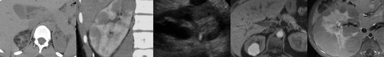
  • Hemangioma has peripheral discontinuous globular enhancement in arterial phase. Progressive centripetal contrast filling and iso- or hyperenhancement in portal venous and late phase
  • Non-hepatocellular malignancy typically demonstrates early (less than 1 minute) and/or marked washout
  • Hemangioma filling may be partial or complete depending on lesion size

To learn more about the Samsung RS85 Prestige ultrasound system, please visit: https://www.bostonimaging.com/rs85-prestige-ultrasound-system-4

See more impressive visuals by filmmaker and freelance motion graphics artist Nick Shaheen: https://www.instagram.com/nickhshaheen

Click the YouTube Community tab or follow on social media for bonus teaching material posted throughout the week!

Spotify: https://spoti.fi/462r0F2
Instagram: https://www.instagram.com/Radquarters/
Facebook: https://www.facebook.com/Radquarters/
X (Twitter): https://twitter.com/Radquarters
Reddit: https://www.reddit.com/user/radiologistHQ/

Ultrasound of Tennis Leg

In this radiology lecture, we review the ultrasound appearance of tennis leg, including medial gastrocnemius and plantaris injury!

Key teaching points include:

  • Tennis leg = Injury to muscles of the calf. Tear of myotendinous junction of medial head of gastrocnemius, rupture of plantaris tendon (less common), in isolation or together
  • Classically described in tennis players, but can occur in various athletic activities (running, skiing) with extension of knee and forced dorsiflexion of ankle. Typically seen in middle-aged, active individuals
  • Clinical: Sudden sharp calf pain with associated popping/snapping sensation followed by tenderness and swelling
  • Gastrocnemius & soleus are pennate muscles. Fascicles attach obliquely to a tendon = Aponeuroses with long length of musculotendinous junction. Feathers converging on a single point
  • Triceps surae muscle = Two headed gastrocnemius, soleus and plantaris. Distal continuation of the gastrocnemius and soleus forms the Achilles tendon
  • Distal medial head of gastrocnemius where tapers over soleus = One of most commonly injured calf structures
  • Medial gastrocnemius tear appears as disrupted tendon fibers at aponeurosis with anechoic/hypoechoic fluid or hemorrhage +/- muscle retraction
  • May see retracted muscle fascicles. Hematoma can dissect between and extends into medial gastrocnemius and soleus muscles
  • Tx: Conservative (self-limiting). Surgical fasciotomy if compartment syndrome
  • Plantaris muscle arises from the posterosuperior aspect of lateral femoral condyle near lateral head origin of gastrocnemius muscle. Medially crosses posterior knee joint in oblique fashion
  • Plantaris continues into calf as a long, thin tendon traveling between medial head of gastrocnemius and soleus muscles. Courses distally at medial aspect of Achilles tendon, usually inserts onto calcaneus. Plantaris is absent in up to 20%
  • Plantaris injury/rupture less common than medial head gastrocnemius tear and typically more proximal in calf (at myotendinous junction)

To learn more about the Samsung RS85 Prestige ultrasound system, please visit: https://www.bostonimaging.com/rs85-prestige-ultrasound-system-4

Click the YouTube Community tab or follow on social media for bonus teaching material posted throughout the week!

Spotify: https://spoti.fi/462r0F2
Instagram: https://www.instagram.com/Radquarters/
Facebook: https://www.facebook.com/Radquarters/
X (Twitter): https://twitter.com/Radquarters
Reddit: https://www.reddit.com/user/radiologistHQ/

Ultrasound of Interstitial Ectopic Pregnancy

In this radiology lecture, we review the ultrasound appearance of interstitial ectopic pregnancy!

Key teaching points include:

  • Interstitial ectopic pregnancies are rare, occurring in proximal (interstitial) portion of fallopian tube within muscle wall of uterus
  • Much less common than tubal ectopic pregnancy occurring in the more distal ampullary and isthmic portions of fallopian tube
  • Interstitial ectopic pregnancies are important because higher morbidity and mortality due to later presentation and risk of life-threatening hemorrhage
  • Abnormally eccentric gestational sac with thin surrounding myometrium: less than 5 mm myometrial thickness highly suspicious
  • “Interstitial line” sign: Thin echogenic line extending from endometrial cavity to ectopic gestational sac. Thought to represent interstitial portion of tube separating the ectopic pregnancy from the endometrium
  • Medical: Systemic MTX, may also be injected into gestational sac
  • Surgery: Cornual wedge resection when ruptured versus hysterectomy
  • Can be confused with angular pregnancy: Rare, intrauterine pregnancy with implantation eccentrically high at the lateral angle of uterine cavity. More medial than interstitial ectopic pregnancies. No interstitial line sign, and greater than 5 mm thickness of overlying myometrial mantle
  • Angular pregnancy can result in normal pregnancy, but increased risk of miscarriage and uterine rupture. Should be followed closely to ensure growth towards endometrial cavity
  • Angular pregnancy is sometimes referred to as a “cornual pregnancy,” but controversial as earliest use of term cornual pregnancy refers to intrauterine implantations in anomalous unicornuate, bicornuate or septate uteri. To avoid confusion, best to specifically describe whether the gestational sac is intrauterine or ectopic

To learn more about the Samsung RS85 Prestige ultrasound system, please visit: https://www.bostonimaging.com/rs85-prestige-ultrasound-system-4

Click the YouTube Community tab or follow on social media for bonus teaching material posted throughout the week!

Spotify: https://spoti.fi/462r0F2
Instagram: https://www.instagram.com/Radquarters/
Facebook: https://www.facebook.com/Radquarters/
X (Twitter): https://twitter.com/Radquarters
Reddit: https://www.reddit.com/user/radiologistHQ/

Ultrasound of Ovarian Serous Cystadenocarcinoma

In this radiology lecture, we review the ultrasound appearance of ovarian serous cystadenocarcinoma!

Key teaching points include:

  • Serous cystadenocarcinoma is the common ovarian malignancy and most common ovarian epithelial tumor
  • High-grade and low-grade types
    Peak incidence 6th-7th decades
  • Ultrasound appearance: Mixed cystic and solid mass with papillary projections and thick septations
  • Elevated CA-125 in greater than 90%
  • Serous tumors are more commonly bilateral than other tumors
  • Four main categories of ovarian neoplasms: Epithelial (most common), germ cell (second most common), sex cord-stromal and metastases
  • Epithelial ovarian tumors are thought to originate outside the ovary (within fallopian tube or endometrium) and involve ovary secondarily
  • Epithelial ovarian tumor types: Serous, mucinous, endometrioid, clear cell and Brenner
  • 60% of epithelial tumors are benign: Unilocular with thin wall or thin septations (less than 3 mm in thickness)
  • 40% of epithelial tumors are malignant or borderline: Papillary projection (distinctive feature of epithelial tumors) with thick, irregular wall or septations (greater than 3 mm in thickness). Can also present as a large soft tissue mass with necrosis. Advanced findings include peritoneal implants, pelvic wall invasion, adenopathy and ascites

To learn more about the Samsung RS85 Prestige ultrasound system, please visit: https://www.bostonimaging.com/rs85-prestige-ultrasound-system-4

Click the YouTube Community tab or follow on social media for bonus teaching material posted throughout the week!

Spotify: https://spoti.fi/462r0F2
Instagram: https://www.instagram.com/Radquarters/
Facebook: https://www.facebook.com/Radquarters/
X (Twitter): https://twitter.com/Radquarters
Reddit: https://www.reddit.com/user/radiologistHQ/

Ultrasound of Sublingual Dermoid Cyst

In this radiology lecture, we review the ultrasound appearance of sublingual dermoid cyst and explain floor of mouth anatomy!

Key teaching points include:

  • The floor of the mouth is a horseshoe-shaped area beneath tongue and in between sides of mandible, inferiorly bounded by mylohyoid muscle, and containing sublingual space (SLS)
  • SLS medial border: Midline genioglossus/geniohyoid muscle complex; SLS inferolateral border: Mylohyoid muscle
  • Anterior margin of hyoglossus muscle projects into posterior SLS
  • Sublingual dermoid cyst is a rare, benign cyst with squamous epithelial lining and contains skin appendages
  • Dermoid and epidermoid cysts are in same family, terminology often used interchangeably, although epidermoid cysts less common and tend to contain fluid contents only
  • Dermoid cyst mean age of presentation late teens to twenties, average age 30
  • Presents as a slowly enlarging neck mass, may cause dysphagia
  • Often round or oval in shape and homogeneously hypoechoic with punctate echogenic foci
  • May have pathognomonic “sack of marbles” appearance
  • Relationship to mylohyoid is key for surgical planning: Intraoral resection for sublingual (above mylohyoid) location, extraoral approach for submental/submandibular (below mylohyoid) location
  • Most cysts are midline
  • DDx: Suprahyoid thyroglossal duct cyst, ranula (simple and diving), abscess and lymphangioma

To learn more about the Samsung RS85 Prestige ultrasound system, please visit: https://www.bostonimaging.com/rs85-prestige-ultrasound-system-4

Click the YouTube Community tab or follow on social media for bonus teaching material posted throughout the week!

Spotify: https://spoti.fi/462r0F2
Instagram: https://www.instagram.com/Radquarters/
Facebook: https://www.facebook.com/Radquarters/
X (Twitter): https://twitter.com/Radquarters
Reddit: https://www.reddit.com/user/radiologistHQ/

Ultrasound of Carpal Tunnel Syndrome

In this radiology lecture, we review the ultrasound appearance of carpal tunnel syndrome!

Key teaching points include:

  • Most common upper extremity entrapment neuropathy. Results from median nerve compression
  • With carpal tunnel syndrome, see hypoechoic enlargement of the median nerve as enters carpal tunnel with flattening of nerve = Notch sign, also volar bowing of flexor retinaculum
  • Median nerve area: Less than 8 mm2 = Normal; 8-12 mm2 = Borderline; greater than 12 mm2 = Abnormal
  • Most accurate to compare nerve area at proximal pronator quadratus muscle and carpal tunnel: Increase of 2 mm2 or more from proximal to distal = 99% sensitive and 100% specific for carpal tunnel syndrome. Measure inside the echogenic epineurium
  • Bifid median nerve: Normal variant in 15% of population, one trunk may take aberrant course through flexor digitorum superficialis musculature, and often associated with persistent median artery between the two trunks
  • Important to recognize persistent median artery pre-operatively because could be damaged during surgery
  • For diagnosis of carpal tunnel syndrome with bifid median nerve: Combined increase of 4 mm2 or more
  • After carpal tunnel release surgery, median nerve may return to normal diameter or remain enlarged regardless of clinical outcome. Retinaculum may appear thickened or disrupted
  • Carpal tunnel syndrome can be caused by extrinsic compression by a mass, ganglion cyst, or tenosynovitis

Reference: Klauser AS, Halpern EJ, De Zordo T, et al. Carpal tunnel syndrome assessment with US: value of additional cross-sectional area measurements of the median nerve in patients versus healthy volunteers. Radiology. 2009;250(1):171-177.

To learn more about the Samsung RS85 Prestige ultrasound system, please visit: https://www.bostonimaging.com/rs85-prestige-ultrasound-system-4

Click the YouTube Community tab or follow on social media for bonus teaching material posted throughout the week!

Spotify: https://spoti.fi/462r0F2
Instagram: https://www.instagram.com/Radquarters/
Facebook: https://www.facebook.com/Radquarters/
Twitter: https://twitter.com/Radquarters
Reddit: https://www.reddit.com/user/radiologistHQ/

Ultrasound of Ganglion Cyst & Wrist Anatomy Review

In this radiology lecture, we review the ultrasound appearance of ganglion cysts while highlighting relevant wrist ultrasound anatomy!

Key teaching points include:

  • Ganglion cysts are viscous, mucin-filled collections lacking a synovial lining
  • Most commonly occur at hand/wrist = Most common wrist mass
  • Location: Dorsum of wrist (60%), frequently adjacent to scapholunate ligament; volar wrist (20%), often between radial artery and flexor carpi radialis tendon; flexor tendon sheath (10%); associated with DIP joint (10%)
  • Grows out of tissues surrounding joint like a balloon on a stalk. May see a pedicle connecting to joint
  • Usually well-defined and multilocular, can be unilocular
  • Hypoechoic to anechoic with posterior acoustic enhancement
  • Noncompressible: Dorsal joint recess and bursal collections will typically collapse with transducer pressure or wrist movement
  • Typically no vascular flow, but septations may have vascularity. May see pulsation artifact from adjacent radial artery
  • Volar cysts can extend towards median nerve and may cause carpal tunnel syndrome
  • May displace or envelop radial artery
  • Tx: Watchful waiting, percutaneous US-guided aspiration and steroid injection, excision
  • Lister’s tubercle is a useful landmark for dorsal wrist anatomy
  • Relevant dorsal extensor tendons (from radial side to ulnar): Compartment 2 = Extensor carpi radialis longus, extensor carpi radialis brevis, Compartment 3 = Extensor pollicis longus (on ulnar side of Lister’s tubercle), Compartment 4 = Extensor digitorum and extensor indicis
  • Flexor carpi radialis overlies the ventral aspect of the scaphoid bone
  • Pisiform and scaphoid bone form the proximal “twin peaks” of the carpal tunnel at the ventral wrist crease
  • Median nerve diameter increase of 2 mm2 or more = Significant compression

To learn more about the Samsung RS85 Prestige ultrasound system, please visit: https://www.bostonimaging.com/rs85-prestige-ultrasound-system-4

Click the YouTube Community tab or follow on social media for bonus teaching material posted throughout the week!

Spotify: https://spoti.fi/462r0F2
Instagram: https://www.instagram.com/Radquarters/
Facebook: https://www.facebook.com/Radquarters/
Twitter: https://twitter.com/Radquarters
Reddit: https://www.reddit.com/user/radiologistHQ/

Radquarters

Radiologist Headquarters has a new name: Radquarters! Same high-yield content, but now with a streamlined name that’s easier to remember.

Click the YouTube Community tab or follow on social media for bonus teaching material posted throughout the week!

Spotify: https://spoti.fi/462r0F2
Instagram: https://www.instagram.com/Radquarters/
Facebook: https://www.facebook.com/Radquarters/
Twitter: https://twitter.com/Radquarters
Reddit: https://www.reddit.com/user/radiologistHQ/

Ultrasound of Epididymitis & Orchitis

In this radiology lecture, we review the ultrasound appearance of acute epididymitis and orchitis!

Key teaching points include:

  • Epididymitis = Inflammation of epididymis. Usually bacterial, most commonly due to retrograde ascent from bladder or prostate.
  • Causative infectious agent varies based on age: Adults younger than 35: Neisseria gonorrhoeae, Chlamydia trachomatis (STDs). Adults older than 35: E. coli & other coliform bacteria.
  • Non-infectious causes of epididymitis: Trauma, repetitive activities such as sports (most common causes in males prior to sexual maturity), torsed appendix testis or appendix epididymis, vasculitis, and medications (amiodarone).
  • Presentation: Gradual onset of scrotal pain, swelling & urinary symptoms. Must exclude testicular torsion (usually more acute onset of pain).
  • Epididymitis US findings: Epididymal enlargement, hyperemia, hypoechogenicity. Hyperemia usually precedes grey scale changes. Infection usually spreads from tail to body and head.
  • 20-30% of epididymitis cases have associated orchitis: Scrotal infection typically starts with epididymis then spreads to testis, scrotal sac, or scrotal wall.
  • Orchitis is less common than and usually secondary to epididymitis. Isolated orchitis uncommon, usually viral (mumps).
  • Orchitis US findings: Testicular enlargement, hyperemia and hypoechogenicity.
  • Complications: Scrotal wall inflammation, complicated hydrocele, pyocele (purulent fluid collection with mass effect), abscess (epididymal, testicular, scrotal wall), testicular ischemia and infarct due to obstructed venous outflow (decreased color Doppler testicular blood flow or reversed testicular diastolic arterial flow).

To learn more about the Samsung RS85 Prestige ultrasound system, please visit: https://www.bostonimaging.com/rs85-prestige-ultrasound-system-4

Click the YouTube Community tab or follow on social media for bonus teaching material posted throughout the week!

Spotify: https://bit.ly/spotify-rhq
Instagram: https://www.instagram.com/radquarters/
Facebook: https://www.facebook.com/radquarters/
Twitter: https://twitter.com/radquarters
Reddit: https://www.reddit.com/user/radiologistHQ/

Ultrasound of Acute Cholecystitis

In this radiology lecture, we review the ultrasound appearance of acute cholecystitis, including gangrenous and emphysematous cholecystitis!

Key teaching points include:

  • Acute cholecystitis = Acute gallbladder inflammation.
  • Most often (95%) caused by an impacted, obstructing gallstone in the cystic duct or gallbladder neck = Acute calculous cholecystitis.
  • Clinically presents as persistent RUQ pain that may radiate to right shoulder, often with N/V and fever.
  • Ultrasound findings of uncomplicated acute cholecystitis: Gallstones, sonographic Murphy sign, gallbladder wall thickening (greater than 3 mm) and edema, gallbladder distention (greater than 4 cm short axis), and pericholecystic fluid.
  • Sonographic Murphy sign = Maximal abdominal tenderness from transducer pressure over gallbladder. PPV of gallstones and a positive sonographic Murphy sign = 92%.
  • Pericholecystic fluid occurs in less than 20% of patients with acute cholecystitis, usually seen in more advanced cases.
  • Gangrenous cholecystitis = Most common complication of acute cholecystitis. Ischemia with necrosis of gallbladder wall. Increased mortality compared to uncomplicated acute cholecystitis.
  • Ultrasound findings of gangrenous cholecystitis: Wall disruption, ulceration, mucosal irregularity, and/or focal bulge, sloughed mucosal membranes, pericholecystic fluid, less likely to have positive Murphy sign, and increased risk of perforation (usually at fundus).
  • Emphysematous cholecystitis = Gallbladder wall necrosis with gas formation in wall and/or lumen. More common in elderly men with underlying diabetes. Higher risk of perforation, rapid progression, and increased mortality compared to uncomplicated acute cholecystitis. Emergent surgical intervention typically required.
  • Ultrasound findings of emphysematous cholecystitis: Bright reflectors from nondependent portions of gallbladder wall, dirty posterior acoustic shadowing, and ring-down artifact. CT can confirm if necessary.

To learn more about the Samsung RS85 Prestige ultrasound system, please visit: https://www.bostonimaging.com/rs85-prestige-ultrasound-system-4

Click the YouTube Community tab or follow on social media for bonus teaching material posted throughout the week!

Spotify: https://bit.ly/spotify-rhq
Instagram: https://www.instagram.com/radiologistHQ/
Facebook: https://www.facebook.com/radiologistHeadQuarters/
Twitter: https://twitter.com/radiologistHQ
Reddit: https://www.reddit.com/user/radiologistHQ/


1 2 3 4 8