Body MRI Physics
- Utility of the FIESTA Pulse Sequence in Body Oncologic Imaging: Review Key Points
“Compared with other steady-state pulse sequences, the FIESTA sequence does not suffer from excessive signal saturation or motion artifacts and offers an excellent image signal-to-noise ratio (SNR). The contrast of the images is not simply T1- or T2-weighted but rather is related to the T2–T1 ratio of the tissue.”
“Although FIESTA images look like gradient-echo images, the signal actually is mostly spin echo and does not depend on T2*, a variant of T2-weighted imaging in which the spins are not refocused to compensate for field inhomogeneities; the SNR of FIESTA images is very high. The contrast of FIESTA images is neither simply T1- nor T2-weighted. Instead, it is determined by the T2–T1 ratio of the tissues. As a result, fluid and fat (which have different T1 and T2 signals but a similar T2–T1 ratio) are usually bright on FIESTA images. On the other hand, muscle, such as myocardium (which has long T1 and short T2 signals), usually is much darker on FIESTA images.”
“With its much shorter acquisition time, FIESTA is less susceptible to respiratory motion artifacts than are T2-weighted fast spin-echo (FSE) or SSFSE sequences.”
“In general, we find that the main advantages of the FIESTA sequence are its motion insensitivity, sharp edge definition, higher contrast when compared with the SSFSE or the SSFSE pulse sequence, and insensitivity to flow void artifacts.”
“Tumors that contain fat tissue, such as adenomas or angiomyolipomas, can be characterized by a decrease in signal intensity with or without applying a fat-suppression technique, such as chemical shift selective saturation, to the FIESTA sequence. The signal drop in a fat-containing tissue on the FIESTA sequence would have similar manifestation as in the in- and out-of-phase pulse sequences.”
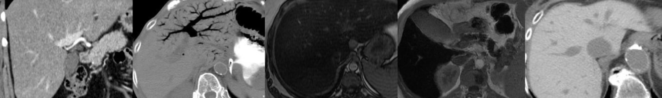
“Clear visualization of the high-signal-intensity peripancreatic vessels on motion-free FIESTA images can be beneficial in evaluating local vascular tumor invasion in the setting of pancreatic neuroendocrine tumors”
“The FIESTA technique is one of the relatively better imaging sequences that can be used to evaluate vascular invasion of renal cell carcinoma without using IV contrast material.”
“The ultrafast FIESTA sequence is capable of depicting the vascular anatomy and its relationship to large primary or metastatic masses in the peritoneal or retroperitoneal cavity, which can contribute to surgical planning. Such comprehensive anatomic detail may not be readily available on any other sequences.”
“Lymph nodes along the drainage pathway of the tumor can be missed on SSFSE images because their signal intensity may be almost identical to that of fat; however, these nodes may be easily identified on a FIESTA sequence because of the edge enhancement capability of the technique or its motion insensitivity that leads to clearer images.”
“An additional benefit of using FIESTA in abdominal imaging is that its high tissue contrast relative to SSFSE makes it easy to screen soft tissue and bone for lesions in an oncology patient.”
“At higher field strengths (e.g., 3 T), the banding artifact may become more severe and appear on axial images at the edge of the coil, mimicking a lipoma. Pulsation artifacts also are often seen on images acquired at 3 T.”
“Although the FIESTA technique can not be used to characterize liver masses, its high SNR, high contrast when compared with SSFSE, short scanning time and motion insensitivity, sharp edge definition, and insensitivity to flow void artifacts make this sequence an excellent adjunct to conventional T1-and T2-weighted sequences for abdominal imaging of cancer patients. In some situations, FIESTA may even be the superior imaging technique.”
“FIESTA is particularly useful in providing clear images of the vascular anatomy in local staging and presurgical planning, particularly for patients with pancreatic, renal, and large retroperitoneal tumors, and in screening for bone and peritoneal tumors.”
- Current MR Imaging Lipid Detection Techniques for Diagnosis of Lesions in the Abdomen and Pelvis